The DR5/TRAILR2/TNFRSF10B Monoclonal Antibody (CAB19043) is a high-quality antibody developed for reliable detection and analysis of target proteins. This antibody, produced in rabbits, is highly specific for human samples and has been validated for use in Western blotting and immunohistochemistry applications.DR5, also known as TNFRSF10B, is a member of the tumor necrosis factor receptor superfamily and is involved in apoptosis, particularly in response to treatment with certain chemotherapeutic drugs.
This antibody is validated for use in WB, IHC-P, IF/ICC, ELISA applications and has demonstrated reactivity against Human, Mouse, Rat samples.
Product Name:
DR5/TRAILR2/TNFRSF10B Monoclonal Antibody
SKU:
CAB19043
Size:
20μL, 100μL
Reactivity:
Human, Mouse, Rat
Clone Number:
ARC0406
Conjugate:
Unconjugated
Immunogen:
Synthetic peptide. This information is considered to be commercially sensitive.
The protein encoded by this gene is a member of the TNF-receptor superfamily, and contains an intracellular death domain. This receptor can be activated by tumor necrosis factor-related apoptosis inducing ligand (TNFSF10/TRAIL/APO-2L), and transduces an apoptosis signal. Studies with FADD-deficient mice suggested that FADD, a death domain containing adaptor protein, is required for the apoptosis mediated by this protein. Two transcript variants encoding different isoforms and one non-coding transcript have been found for this gene.
Purification Method
Affinity purification
Gene ID
8795
RRID
AB_2862536
Buffer Information
Store at -20℃. Avoid freeze / thaw cycles. Buffer: PBS containing 50% glycerol and 0.05% BSA, preserved with proclin300 or sodium azide, pH 7.3.
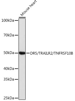
Western blot analysis of lysates from Mouse heart, using DR5/TRAILR2/TNFRSF10B Rabbit mAb (CAB19043) at 1:1000 dilution. Secondary antibody: HRP-conjugated Goat anti-Rabbit IgG (H+L) (CABS014) at 1:10000 dilution. Lysates/proteins: 25μg per lane. Blocking buffer: 3% nonfat dry milk in TBST. Detection: ECL Basic Kit (AbGn00020). Exposure time: 1s.
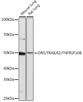
Western blot analysis of various lysates using DR5/TRAILR2/TNFRSF10B Rabbit mAb (CAB19043) at 1:1000 dilution. Secondary antibody: HRP-conjugated Goat anti-Rabbit IgG (H+L) (CABS014) at 1:10000 dilution. Lysates/proteins: 25μg per lane. Blocking buffer: 3% nonfat dry milk in TBST. Detection: ECL Basic Kit (AbGn00020). Exposure time: 3min.
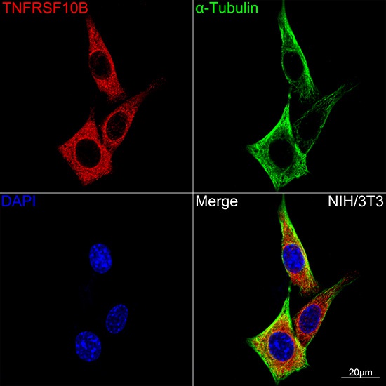
Confocal imaging of NIH-3T3 cells using DR5/TRAILR2/TNFRSF10B Rabbit mAb (CAB19043,dilution 1:100)(Red). The cells were counterstained with α-Tubulin Mouse mAb (AC012,dilution 1:400) (Green). DAPI was used for nuclear staining (blue). Objective: 100x.


")
")
")
