The DUSP6 Monoclonal Antibody (CAB0133) is a high-quality antibody developed for reliable detection and analysis of target proteins. This antibody, generated in rabbits, exhibits high specificity and sensitivity for detecting DUSP6 in human samples, making it ideal for Western blot applications. By binding to the DUSP6 protein, researchers can accurately analyze and quantify its expression in various cell types.
This antibody is validated for use in WB, IHC-P, IP, ELISA applications and has demonstrated reactivity against Human, Mouse, Rat samples.
Product Name:
DUSP6 Monoclonal Antibody
SKU:
CAB0133
Size:
20μL, 100μL
Reactivity:
Human, Mouse, Rat
Clone Number:
ARC0237
Conjugate:
Unconjugated
Immunogen:
Synthetic peptide. This information is considered to be commercially sensitive.
0.5μg-4μg antibody for 200μg-400μg extracts of whole cells
IHC-P
1:200 - 1:800
ELISA
Recommended starting concentration is 1 μg/mL. Please optimize the concentration based on your specific assay requirements.For high-ratio antibody dilutions (≥1:10000),a sequential dilution method is strongly recommended to ensure measurement accuracy.
Synonyms:
HH19, MKP3, PYST1, DUSP6
Positive Sample:
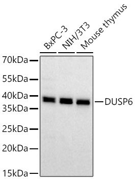
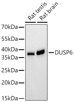
Rat testis, Rat brain, BxPC-3, NIH/3T3, Mouse thymus
Cellular Localization:
Cytoplasm.
Calculated MW:
42kDa
Observed MW:
42kDa
The protein encoded by this gene is a member of the dual specificity protein phosphatase subfamily. These phosphatases inactivate their target kinases by dephosphorylating both the phosphoserine/threonine and phosphotyrosine residues. They negatively regulate members of the mitogen-activated protein (MAP) kinase superfamily (MAPK/ERK, SAPK/JNK, p38), which are associated with cellular proliferation and differentiation. Different members of the family of dual specificity phosphatases show distinct substrate specificities for various MAP kinases, different tissue distribution and subcellular localization, and different modes of inducibility of their expression by extracellular stimuli. This gene product inactivates ERK2, is expressed in a variety of tissues with the highest levels in heart and pancreas, and unlike most other members of this family, is localized in the cytoplasm. Mutations in this gene have been associated with congenital hypogonadotropic hypogonadism. Alternatively spliced transcript variants have been found for this gene.
Purification Method
Affinity purification
Gene ID
1848
RRID
AB_2861454
Buffer Information
Store at -20℃. Avoid freeze / thaw cycles. Buffer: PBS containing 50% glycerol and 0.05% BSA, preserved with proclin300 or sodium azide, pH 7.3.
Western blot analysis of various lysates using DUSP6 Rabbit mAb (CAB0133) at 1:1000 dilution incubated at room temperature for 1.5 hours. Secondary antibody: HRP-conjugated Goat anti-Rabbit IgG (H+L) (CABS014) at 1:10000 dilution. Lysates/proteins: 25 μg per lane. Blocking buffer: 3% nonfat dry milk in TBST. Detection: ECL Basic Kit (AbGn00020). Exposure time: 45s.
Western blot analysis of various lysates using DUSP6 Rabbit mAb (CAB0133) at 1:1000 dilution incubated at room temperature for 1.5 hours. Secondary antibody: HRP-conjugated Goat anti-Rabbit IgG (H+L) (CABS014) at 1:10000 dilution. Lysates/proteins: 25 μg per lane. Blocking buffer: 3% nonfat dry milk in TBST. Detection: ECL Basic Kit (AbGn00020). Exposure time: 90s.
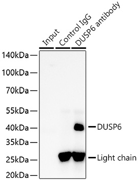
Immunoprecipitation of DUSP6 from 300 µg extracts of Hep G2 cells was performed using 2 µg of DUSP6 Rabbit mAb (CAB0133). Rabbit Control IgG (AC005) was used to precipitate the Control IgG sample. IP samples were eluted with 1x Laemmli Buffer. The Input lane represents 10% of the total input. Western blot analysis of immunoprecipitates was conducted using DUSP6 Rabbit mAb (CAB0133) at a dilution of 1:10000.