The ECI1 Antibody (CAB1211) is a high-quality antibody developed for reliable detection and analysis of target proteins. This antibody, produced in rabbits, is highly specific for detecting ECI1 in human samples and is validated for use in Western blot applications. By binding to the ECI1 protein, this antibody allows for precise detection and analysis of ECI1 expression in various cell types, making it an essential tool for studies in lipid metabolism, cell signaling, and related fields.
This antibody is validated for use in WB, IHC-P, IF/ICC, ELISA applications and has demonstrated reactivity against Human, Mouse samples.
Product Name:
ECI1 Antibody
SKU:
CAB1211
Size:
20μL, 100μL
Reactivity:
Human, Mouse
Conjugate:
Unconjugated
Immunogen:
Recombinant protein (or fragment).This information is considered to be commercially sensitive.
Recommended starting concentration is 1 μg/mL. Please optimize the concentration based on your specific assay requirements.
Synonyms:
DCI, ECI1
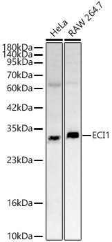
Positive Sample:
HeLa, RAW 264.7
Cellular Localization:
Mitochondrion Matrix.
Calculated MW:
33kDa
Observed MW:
33kDa
This gene encodes a member of the hydratase/isomerase superfamily. The protein encoded is a key mitochondrial enzyme involved in beta-oxidation of unsaturated fatty acids. It catalyzes the transformation of 3-cis and 3-trans-enoyl-CoA esters arising during the stepwise degradation of cis-, mono-, and polyunsaturated fatty acids to the 2-trans-enoyl-CoA intermediates. Alternatively spliced transcript variants have been described.
Purification Method
Affinity purification
Gene ID
1632
RRID
AB_2759001
Buffer Information
Store at -20℃. Avoid freeze / thaw cycles. Buffer: PBS containing 50% glycerol, preserved with proclin300 or sodium azide, pH 7.3.
Western blot analysis of various lysates using ECI1 Rabbit pAb (CAB1211) at 1:2000 dilution. Secondary antibody: HRP-conjugated Goat anti-Rabbit IgG (H+L) (CABS014) at 1:10000 dilution. Lysates / proteins: 25 μg per lane. Blocking buffer: 3 % nonfat dry milk in TBST. Detection: ECL Basic Kit (AbGn00020). Exposure time: 45s.
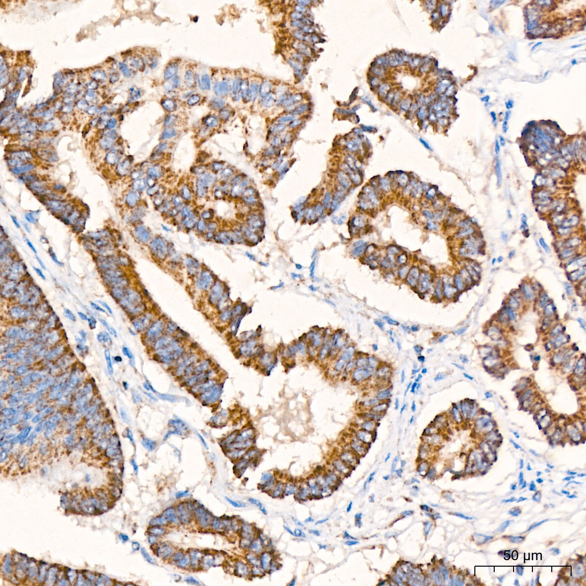
Immunohistochemistry analysis of paraffin-embedded Human colon carcinoma tissue using ECI1 Rabbit pAb (CAB1211) at a dilution of 1:200 (40x lens). High pressure antigen retrieval was performed with 0.01 M citrate buffer (pH 6.0) prior to IHC staining.