The ECM1 Antibody (CAB16368) is a high-quality antibody developed for reliable detection and analysis of target proteins. The antibody, produced in rabbits, shows high reactivity with human samples and is validated for use in various applications including Western blot and immunohistochemistry.ECM1, or extracellular matrix protein 1, is essential for maintaining the structural integrity of tissues and plays a crucial role in processes such as wound healing and tissue remodeling. Dysregulation of ECM1 has been implicated in various diseases including cancer and fibrotic disorders, making it a promising target for therapeutic interventions.
This antibody is validated for use in WB, IF/ICC, ELISA applications and has demonstrated reactivity against Human, Mouse samples.
Product Name:
ECM1 Antibody
SKU:
CAB16368
Size:
20μL, 100μL
Reactivity:
Human, Mouse
Conjugate:
Unconjugated
Immunogen:
Recombinant protein (or fragment).This information is considered to be commercially sensitive.
This gene encodes a soluble protein that is involved in endochondral bone formation, angiogenesis, and tumor biology. It also interacts with a variety of extracellular and structural proteins, contributing to the maintenance of skin integrity and homeostasis. Mutations in this gene are associated with lipoid proteinosis disorder (also known as hyalinosis cutis et mucosae or Urbach-Wiethe disease) that is characterized by generalized thickening of skin, mucosae and certain viscera. Alternatively spliced transcript variants encoding distinct isoforms have been described for this gene.
Purification Method
Affinity purification
Gene ID
1893
RRID
AB_2769259
Buffer Information
Store at -20℃. Avoid freeze / thaw cycles. Buffer: PBS containing 50% glycerol, preserved with proclin300 or sodium azide, pH 7.3.
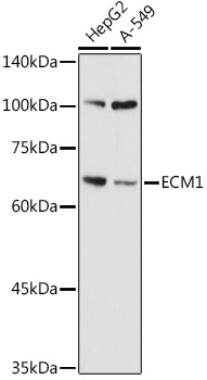
Western blot analysis of various lysates using ECM1 Rabbit pAb (CAB16368) at 1:1000 dilution. Secondary antibody: HRP-conjugated Goat anti-Rabbit IgG (H+L) (CABS014) at 1:10000 dilution. Lysates/proteins: 25μg per lane. Blocking buffer: 3% nonfat dry milk in TBST. Detection: ECL Basic Kit (AbGn00020). Exposure time: 10s.
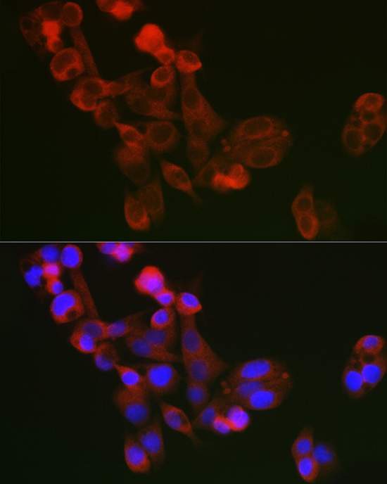
Immunofluorescence analysis of A375 cells using ECM1 Rabbit pAb (CAB16368) at dilution of 1:100 (40x lens). Secondary antibody: Cy3-conjugated Goat anti-Rabbit IgG (H+L) (CABS007) at 1:500 dilution. Blue: DAPI for nuclear staining.