The EEA1 Antibody (CAB0592) is a high-quality antibody developed for reliable detection and analysis of target proteins. This antibody, raised in rabbits, is highly specific to the Early Endosome Antigen 1 (EEA1) protein and has been rigorously validated for use in various applications, including immunofluorescence and immunohistochemistry.EEA1 is a key component of the early endosome compartment, playing a crucial role in sorting and trafficking of endocytic vesicles. Its involvement in membrane fusion events and signaling cascades makes it a target of interest in studies of cellular transport processes and intracellular trafficking pathways. The EEA1 Polyclonal Antibody enables precise detection and visualization of EEA1 protein in different cell types and tissues, providing valuable insights into endosomal dynamics and organelle interactions.
This antibody is validated for use in WB, IHC-P, ELISA applications and has demonstrated reactivity against Human, Mouse, Rat samples.
Product Name:
EEA1 Antibody
SKU:
CAB0592
Size:
20μL, 100μL
Reactivity:
Human, Mouse, Rat
Conjugate:
Unconjugated
Immunogen:
Recombinant protein (or fragment).This information is considered to be commercially sensitive.
Recommended starting concentration is 1 μg/mL. Please optimize the concentration based on your specific assay requirements.
Synonyms:
MST105, ZFYVE2, MSTP105, EEA1
Positive Sample:
U-87MG, HT-1080, LO2, Mouse kidney, Mouse lung, Rat brain
Cellular Localization:
Cytoplasm, Early Endosome Membrane, Peripheral Membrane Protein.
Calculated MW:
162kDa
Observed MW:
175kDa
Enables 1-phosphatidylinositol binding activity; GTP-dependent protein binding activity; and protein homodimerization activity. Involved in endocytosis; vesicle fusion; and viral RNA genome replication. Located in cytosol and early endosome. Is extrinsic component of plasma membrane.
Purification Method
Affinity purification
Gene ID
8411
RRID
AB_2757284
Buffer Information
Store at -20℃. Avoid freeze / thaw cycles. Buffer: PBS containing 50% glycerol, preserved with proclin300 or sodium azide, pH 7.3.
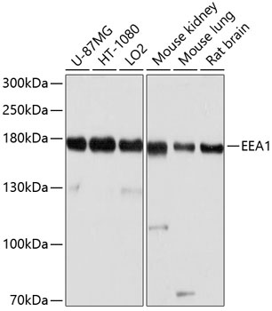
Western blot analysis of various lysates using EEA1 Rabbit pAb (CAB0592) at 1:1000 dilution. Secondary antibody: HRP-conjugated Goat anti-Rabbit IgG (H+L) (CABS014) at 1:10000 dilution. Lysates/proteins: 25μg per lane. Blocking buffer: 3% nonfat dry milk in TBST. Detection: ECL Basic Kit (AbGn00020). Exposure time: 5s.
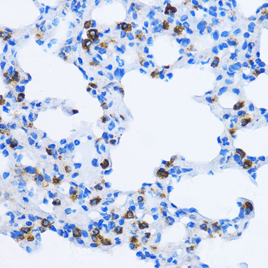
Immunohistochemistry analysis of paraffin-embedded Rat lung using EEA1 Rabbit pAb (CAB0592) at dilution of 1:100 (40x lens). Microwave antigen retrieval performed with 0.01M PBS Buffer (pH 7.2) prior to IHC staining.
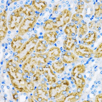
Immunohistochemistry analysis of paraffin-embedded Mouse kidney using EEA1 Rabbit pAb (CAB0592) at dilution of 1:100 (40x lens). Microwave antigen retrieval performed with 0.01M PBS Buffer (pH 7.2) prior to IHC staining.