The [KO Validated] EGFR Antibody (CAB2069) is a high-quality antibody developed for reliable detection and analysis of target proteins. This antibody, generated in rabbits, exhibits high reactivity with human samples and has been validated for use in Western blot applications. It specifically binds to the EGFR protein, enabling accurate detection and analysis in a variety of cell types, making it ideal for studies in cancer biology and signal transduction pathways.
This antibody is validated for use in WB, IP, ELISA applications and has demonstrated reactivity against Human, Mouse, Rat samples.
Product Name:
[KO Validated] EGFR Antibody
SKU:
CAB2069
Size:
20μL, 100μL
Reactivity:
Human, Mouse, Rat
Conjugate:
Unconjugated
Immunogen:
Synthetic peptide. This information is considered to be commercially sensitive.
HeLa, Mouse liver, Rat brain, Rat liver, Rat liver, Rat liver
Cellular Localization:
Cell Membrane, Endoplasmic Reticulum Membrane, Endosome, Endosome Membrane, Golgi Apparatus Membrane, Nucleus Membrane, Nucleus, Secreted, Single-Pass Type I Membrane Protein.
Calculated MW:
134kDa
Observed MW:
175kDa//175kDa/175kDa
The protein encoded by this gene is a transmembrane glycoprotein that is a member of the protein kinase superfamily. This protein is a receptor for members of the epidermal growth factor family. EGFR is a cell surface protein that binds to epidermal growth factor, thus inducing receptor dimerization and tyrosine autophosphorylation leading to cell proliferation. Mutations in this gene are associated with lung cancer. EGFR is a component of the cytokine storm which contributes to a severe form of Coronavirus Disease 2019 (COVID-19) resulting from infection with severe acute respiratory syndrome coronavirus-2 (SARS-CoV-2).
Purification Method
Affinity purification
Gene ID
1956
RRID
AB_2764092
Buffer Information
Store at -20℃. Avoid freeze / thaw cycles. Buffer: PBS containing 50% glycerol, preserved with proclin300 or sodium azide, pH 7.3.
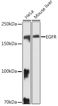
Western blot analysis of various lysates using EGFR Rabbit pAb (CAB2069) at 1:1000 dilution. Secondary antibody: HRP-conjugated Goat anti-Rabbit IgG (H+L) (CABS014) at 1:10000 dilution. Lysates/proteins: 25μg per lane. Blocking buffer: 3% nonfat dry milk in TBST. Detection: ECL Basic Kit (AbGn00020). Exposure time: 10s.
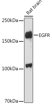
Western blot analysis of lysates from Rat brain, using EGFR Rabbit pAb (CAB2069) at 1:1000 dilution. Secondary antibody: HRP-conjugated Goat anti-Rabbit IgG (H+L) (CABS014) at 1:10000 dilution. Lysates/proteins: 25μg per lane. Blocking buffer: 3% nonfat dry milk in TBST. Detection: ECL Enhanced Kit (AbGn00021). Exposure time: 180s.
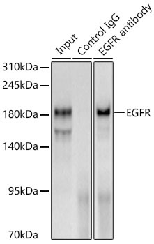
Immunoprecipitation of EGFR from 600 µg extracts of Rat liver tissue was performed using 3 µg of EGFR Rabbit pAb (CAB2069). Rabbit IgG isotype control (AC042) was used to precipitate the Control IgG sample. IP samples were eluted with 1X Laemmli Buffer. The Input lane represents 10% of the total input. Western blot analysis of immunoprecipitates was conducted using EGFR Rabbit pAb (CAB2069) at a dilution of 1:1000.
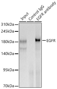
Immunoprecipitation of EGFR from 600 µg extracts of Rat liver tissue was performed using 3 µg of EGFR Rabbit pAb (CAB2069). Rabbit IgG isotype control (AC042) was used to precipitate the Control IgG sample. IP samples were eluted with 1X Laemmli Buffer. The Input lane represents 10% of the total input. Western blot analysis of immunoprecipitates was conducted using EGFR Rabbit pAb (CAB2069) at a dilution of 1:1000.