The EIF3C Antibody (CAB7022) is a high-quality antibody developed for reliable detection and analysis of target proteins. This antibody, produced in rabbits, has high reactivity with human samples and has been validated for use in Western blot applications. EIF3C plays a crucial role in protein synthesis by aiding in the initiation of mRNA translation. Dysregulation of EIF3C has been implicated in various diseases, including cancer, making it a valuable target for research in oncology and other fields.
This antibody is validated for use in WB, IHC-P, IF/ICC, ELISA applications and has demonstrated reactivity against Human, Mouse, Rat samples.
Product Name:
EIF3C Antibody
SKU:
CAB7022
Size:
20μL, 100μL
Reactivity:
Human, Mouse, Rat
Conjugate:
Unconjugated
Immunogen:
Recombinant protein (or fragment).This information is considered to be commercially sensitive.
Recommended starting concentration is 1 μg/mL. Please optimize the concentration based on your specific assay requirements.
Synonyms:
EIF3CL, EIF3S8, eIF3-p110, EIF3C
Positive Sample:
MCF7, HL-60, HepG2, SW620, THP-1, Mouse testis
Cellular Localization:
Cytoplasm.
Calculated MW:
106kDa
Observed MW:
105kDa
Enables ribosome binding activity. Contributes to translation initiation factor activity. Involved in positive regulation of mRNA binding activity; positive regulation of translation; and translational initiation. Part of eukaryotic translation initiation factor 3 complex.
Purification Method
Affinity purification
Gene ID
8663
RRID
AB_2767578
Buffer Information
Store at -20℃. Avoid freeze / thaw cycles. Buffer: PBS containing 50% glycerol, preserved with proclin300 or sodium azide, pH 7.3.
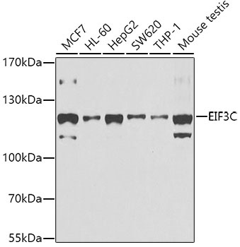
Western blot analysis of various lysates using EIF3C Rabbit pAb (CAB7022) at 1:1000 dilution. Secondary antibody: HRP-conjugated Goat anti-Rabbit IgG (H+L) (CABS014) at 1:10000 dilution. Lysates/proteins: 25μg per lane. Blocking buffer: 3% nonfat dry milk in TBST. Detection: ECL Basic Kit (AbGn00020). Exposure time: 30s.
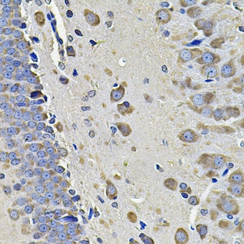
Immunohistochemistry analysis of paraffin-embedded Rat brain using EIF3C Rabbit pAb (CAB7022) at dilution of 1:100 (40x lens). Microwave antigen retrieval performed with 0.01M PBS Buffer (pH 7.2) prior to IHC staining.
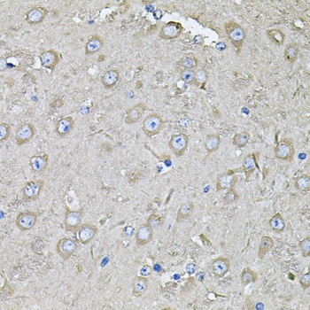
Immunohistochemistry analysis of paraffin-embedded Mouse brain using EIF3C Rabbit pAb (CAB7022) at dilution of 1:100 (40x lens). Microwave antigen retrieval performed with 0.01M PBS Buffer (pH 7.2) prior to IHC staining.
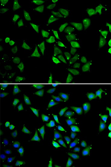
Immunofluorescence analysis of U2OS cells using EIF3C Rabbit pAb (CAB7022). Secondary antibody: Cy3-conjugated Goat anti-Rabbit IgG (H+L) (CABS007) at 1:500 dilution. Blue: DAPI for nuclear staining.