The FAH Antibody (CAB13492) is a high-quality antibody developed for reliable detection and analysis of target proteins. This antibody, produced in rabbits, is highly specific for human samples and has been validated for use in Western blot applications.FAH deficiency is a rare genetic disorder that results in the accumulation of toxic metabolites leading to liver and kidney damage. Research into FAH and its role in tyrosine metabolism is essential for understanding the pathophysiology of this disorder and developing treatments.
This antibody is validated for use in WB, ELISA applications and has demonstrated reactivity against Human, Mouse, Rat samples.
Product Name:
FAH Antibody
SKU:
CAB13492
Size:
20μL, 100μL
Reactivity:
Human, Mouse, Rat
Conjugate:
Unconjugated
Immunogen:
Recombinant protein (or fragment).This information is considered to be commercially sensitive.
Recommended starting concentration is 1 μg/mL. Please optimize the concentration based on your specific assay requirements.
Synonyms:
FAH
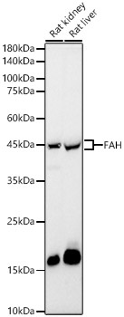
Positive Sample:
Rat kidney, Rat liver
Cellular Localization:
Cytosol, Extracellular Exosome.
Calculated MW:
46kDa
Observed MW:
40-45kDa
Predicted to enable fumarylacetoacetase activity. Predicted to be involved in L-phenylalanine catabolic process; homogentisate catabolic process; and tyrosine catabolic process. Predicted to act upstream of or within arginine catabolic process. Located in extracellular exosome. Implicated in tyrosinemia type I.
Purification Method
Affinity purification
Gene ID
2184
RRID
AB_2760355
Buffer Information
Store at -20℃. Avoid freeze / thaw cycles. Buffer: PBS containing 50% glycerol, preserved with proclin300 or sodium azide, pH 7.3.
Western blot analysis of various lysates, using FAH Rabbit pAb (CAB13492) at 1:7000 dilution. Secondary antibody: HRP-conjugated Goat anti-Rabbit IgG (H+L) (CABS014) at 1:10000 dilution. Lysates/proteins: 25μg per lane. Blocking buffer: 3% nonfat dry milk in TBST. Detection: ECL Basic Kit (AbGn00020). Exposure time: 60s.