FAM83D Antibody is a premium polyclonal that offers outstanding performance and reliability for demanding research applications. Rigorously validated for ELISA, WB, IF, this antibody ensures consistent, reproducible results across multiple experimental platforms. Demonstrates excellent reactivity with Human samples, providing researchers with confidence in cross-species compatibility. Conveniently packaged in 50ug format to meet your experimental needs. For optimal performance, store at -20°C or -80°C and maintains stability for 12 months. Backed by rigorous quality control testing to ensure superior performance in your critical research applications.
Product Name:
FAM83D Antibody
SKU:
PACO60412
Size:
50μg
Isotype:
IgG
Host Species:
Rabbit
Reactivity:
Human
Immunogen:
Recombinant Human Protein FAM83D protein (339-470AA)
Immunogen Species:
Homo sapiens (Human)
Uniprot No:
Q9H4H8
Form:
Liquid
Tested Applications:
ELISAWBIHC
Recommended Dilution:
Application
Recommended Dilution
WB
1:500-1:5000
IHC
1:100-1:300
Synonyms:
2310007D09Rik antibody, BB104611 antibody, C20orf129 antibody, CHICA antibody, dJ616B8.3 antibody, FA83D antibody, FA83D_HUMAN antibody, FAM83D antibody, Family with sequence similarity 83, member D antibody, FLJ38341 antibody, MGC92947 antibody, Protein FAM83D antibody, RGD1565583 antibody, RP23-119H5.1 antibody, Spindle protein CHICA antibody
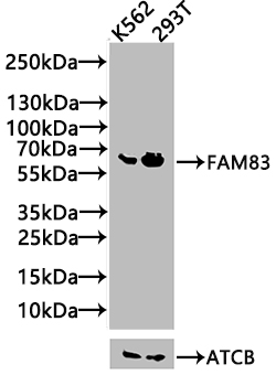
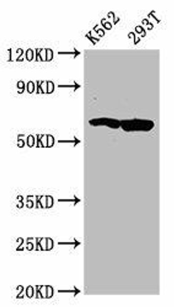
Western Blot Positive WB detected in: K562 whole cell lysate(20µg), 293T whole cell lysate(20µg) All lanes: FAM83D antibody at 1:1000 Secondary Goat polyclonal to rabbit IgG at 1/20000 dilution Predicted band size: 63, 65 kDa Observed band size: 65 kDa Exposure time: 120s
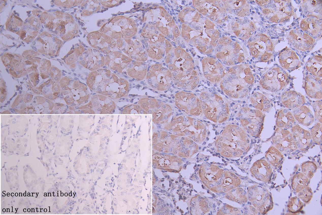
IHC image of PACO60412 diluted at 1:300 and staining in paraffin-embedded human stomach tissue performed on a Leica BondTM system. After dewaxing and hydration, antigen retrieval was mediated by high pressure in a citrate buffer (pH 6.0). Section was blocked with 10% normal goat serum 30min at RT. Then primary antibody (1% BSA) was incubated at 4°C overnight. The primary is detected by a Goat anti-rabbit polymer IgG labeled by HRP and visualized using 0.05% DAB. Secondary antibody only control: uses 1% BSA instead of primary antibody
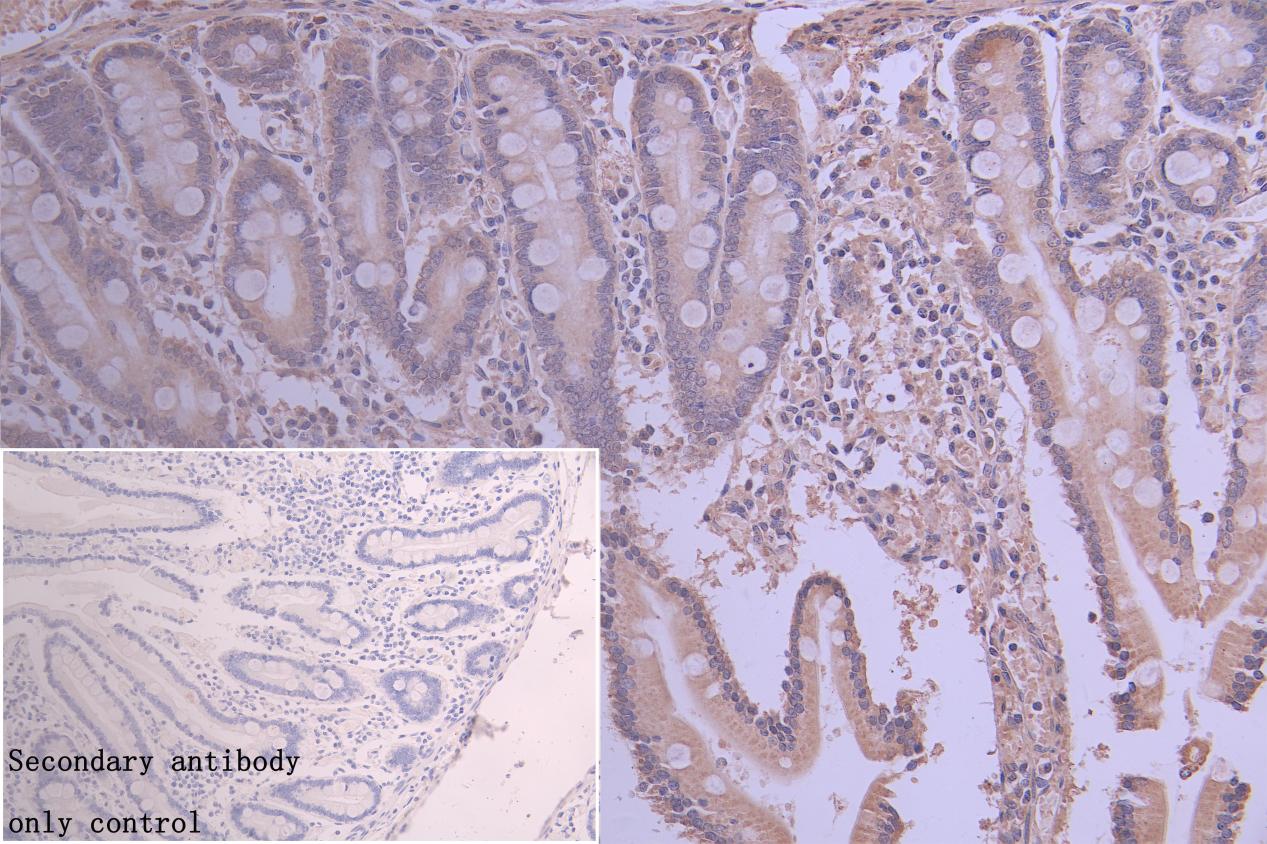
IHC image of PACO60412 diluted at 1:300 and staining in paraffin-embedded human gastric tissue performed on a Leica BondTM system. After dewaxing and hydration, antigen retrieval was mediated by high pressure in a citrate buffer (pH 6.0). Section was blocked with 10% normal goat serum 30min at RT. Then primary antibody (1% BSA) was incubated at 4°C overnight. The primary is detected by a Goat anti-rabbit polymer IgG labeled by HRP and visualized using 0.05% DAB. Secondary antibody only control: uses 1% BSA instead of primary antibody