The FCGR1A Antibody (CAB1197) is a high-quality antibody developed for reliable detection and analysis of target proteins. This antibody, produced in rabbits, has high reactivity with human samples and has been validated for use in Western blot applications. By targeting the FCGR1A protein, this antibody allows for precise detection and analysis in a variety of cell types, making it ideal for studies in immunology and cancer research.FCGR1A, also known as Fc gamma receptor 1A, is involved in immune regulation by mediating the interaction between immune cells and antibodies. Its role in modulating immune responses makes it a promising target for research into diseases such as cancer, autoimmune disorders, and inflammatory conditions.
This antibody is validated for use in WB, IF/ICC, ELISA applications and has demonstrated reactivity against Human, Mouse, Rat samples.
Product Name:
FCGR1A Antibody
SKU:
CAB1197
Size:
20μL, 100μL
Reactivity:
Human, Mouse, Rat
Conjugate:
Unconjugated
Immunogen:
Recombinant protein (or fragment).This information is considered to be commercially sensitive.
Recommended starting concentration is 1 μg/mL. Please optimize the concentration based on your specific assay requirements.
Synonyms:
CD64, FCG1, FCRI, CD64A, FCGR1, IGFR1, FcgammaRI
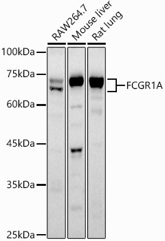
Positive Sample:
RAW264.7, Mouse liver, Rat lung
Cellular Localization:
Cell Membrane, Single-Pass Type I Membrane Protein.
Calculated MW:
43kDa
Observed MW:
65-70kDa
This gene encodes a protein that plays an important role in the immune response. This protein is a high-affinity Fc-gamma receptor. The gene is one of three related gene family members located on chromosome 1.
Purification Method
Affinity purification
Gene ID
2209
RRID
AB_2758903
Buffer Information
Store at -20℃. Avoid freeze / thaw cycles. Buffer: PBS containing 50% glycerol, preserved with proclin300 or sodium azide, pH 7.3.
Western blot analysis of various lysates using CD64 Rabbit pAb (CAB1197) at 1:1000 dilution. Secondary antibody: HRP-conjugated Goat anti-Rabbit IgG (H+L) (CABS014) at 1:10000 dilution. Lysates/proteins: 25μg per lane. Blocking buffer: 3% nonfat dry milk in TBST. Detection: ECL Basic Kit (AbGn00020). Exposure time: 90s.
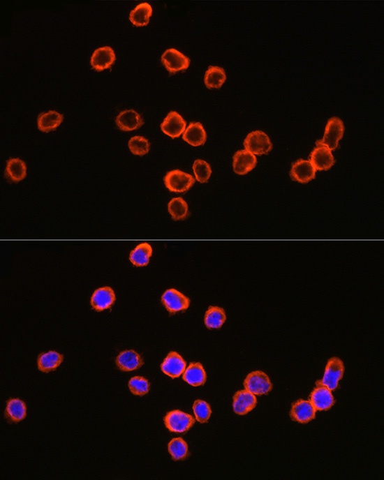
Immunofluorescence analysis of THP-1 cells using CD64 Rabbit pAb (CAB1197) at dilution of 1:100 (40x lens). Secondary antibody: Cy3-conjugated Goat anti-Rabbit IgG (H+L) (CABS007) at 1:500 dilution. Blue: DAPI for nuclear staining.