The FHIT Monoclonal Antibody (CAB9072) is a high-quality antibody developed for reliable detection and analysis of target proteins. This antibody, developed in rabbits, shows high specificity and sensitivity towards FHIT in human samples, making it a reliable choice for Western blot applications. By binding to the FHIT protein, this antibody enables precise detection and analysis in different cell types, offering valuable insights for cancer research and drug development efforts.
This antibody is validated for use in WB, ELISA applications and has demonstrated reactivity against Human, Mouse, Rat samples.
Product Name:
FHIT Monoclonal Antibody
SKU:
CAB9072
Size:
20μL, 100μL
Reactivity:
Human, Mouse, Rat
Clone Number:
ARC1401
Conjugate:
Unconjugated
Immunogen:
Synthetic peptide. This information is considered to be commercially sensitive.
Recommended starting concentration is 1 μg/mL. Please optimize the concentration based on your specific assay requirements.
Synonyms:
FRA3B, AP3Aase, FHIT
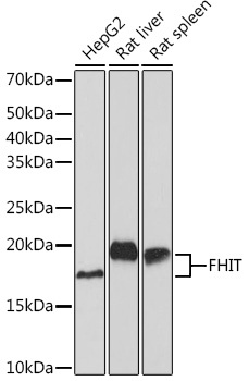
Positive Sample:
HepG2, Rat liver, Rat spleen
Cellular Localization:
Cytoplasm, Mitochondrion, Nucleus.
Calculated MW:
17kDa
Observed MW:
17kDa
The protein encoded by this gene is a P1-P3-bis(5'-adenosyl) triphosphate hydrolase involved in purine metabolism. This gene encompasses the common fragile site FRA3B on chromosome 3, where carcinogen-induced damage can lead to translocations and aberrant transcripts. In fact, aberrant transcripts from this gene have been found in about half of all esophageal, stomach, and colon carcinomas. The encoded protein is also a tumor suppressor, as loss of its activity results in replication stress and DNA damage.
Purification Method
Affinity purification
Gene ID
2272
RRID
AB_2863651
Buffer Information
Store at -20℃. Avoid freeze / thaw cycles. Buffer: PBS containing 50% glycerol and 0.05% BSA, preserved with proclin300 or sodium azide, pH 7.3.
Western blot analysis of various lysates using FHIT Rabbit mAb (CAB9072) at 1:1000 dilution. Secondary antibody: HRP-conjugated Goat anti-Rabbit IgG (H+L) (CABS014) at 1:10000 dilution. Lysates/proteins: 25μg per lane. Blocking buffer: 3% nonfat dry milk in TBST. Detection: ECL Basic Kit (AbGn00020). Exposure time: 60s.