The GALC Antibody (CAB3873) is a high-quality antibody developed for reliable detection and analysis of target proteins. This antibody, produced in rabbits, exhibits high reactivity with human samples and has been validated for use in various applications such as Western blotting. By binding specifically to the GALC protein, this antibody allows for accurate detection and analysis in a variety of cell types, making it a versatile tool for research in biochemistry and neurology.GALC, also known as galactocerebrosidase, is responsible for breaking down specific fats in the body, particularly within the nervous system.
This antibody is validated for use in WB, IHC-P, IF/ICC, ELISA applications and has demonstrated reactivity against Human, Mouse, Rat samples.
Product Name:
GALC Antibody
SKU:
CAB3873
Size:
20μL, 100μL
Reactivity:
Human, Mouse, Rat
Conjugate:
Unconjugated
Immunogen:
Recombinant protein (or fragment).This information is considered to be commercially sensitive.
Recommended starting concentration is 1 μg/mL. Please optimize the concentration based on your specific assay requirements.
Synonyms:
GALC
Positive Sample:
DU145, 293T, HepG2, A375, Mouse brain, Rat testis
Cellular Localization:
Lysosome.
Calculated MW:
77kDa
Observed MW:
77kDa
This gene encodes a lysosomal protein which hydrolyzes the galactose ester bonds of galactosylceramide, galactosylsphingosine, lactosylceramide, and monogalactosyldiglyceride. Mutations in this gene have been associated with Krabbe disease, also known as globoid cell leukodystrophy. Alternate transcriptional splice variants, encoding different isoforms, have been characterized.
Purification Method
Affinity purification
Gene ID
2581
RRID
AB_2765354
Buffer Information
Store at -20℃. Avoid freeze / thaw cycles. Buffer: PBS containing 50% glycerol, preserved with proclin300 or sodium azide, pH 7.3.
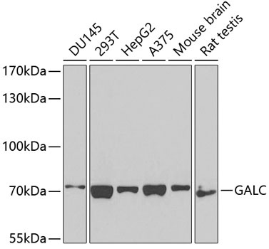
Western blot analysis of various lysates using GALC Rabbit pAb (CAB3873) at 1:1000 dilution. Secondary antibody: HRP-conjugated Goat anti-Rabbit IgG (H+L) (CABS014) at 1:10000 dilution. Lysates/proteins: 25μg per lane. Blocking buffer: 3% nonfat dry milk in TBST. Detection: ECL Basic Kit (AbGn00020). Exposure time: 90s.
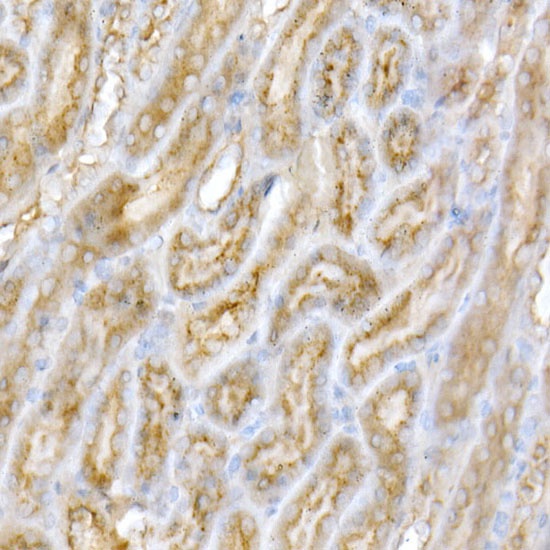
Immunohistochemistry analysis of paraffin-embedded Human kidney using GALC Rabbit pAb (CAB3873) at dilution of 1:200 (40x lens). High pressure antigen retrieval performed with 0.01M Citrate buffer (pH 6.0) prior to IHC staining.