The GLUD2 Antibody (CAB6604) is a high-quality antibody developed for reliable detection and analysis of target proteins. This antibody, produced in rabbits, exhibits high reactivity with human samples and has been rigorously validated for Western blot applications.GLUD2 is known to play a crucial role in the metabolism of glutamate, a key neurotransmitter in the central nervous system. Dysregulation of GLUD2 activity has been implicated in various neurological disorders, making it a target of interest for researchers studying conditions such as epilepsy, Parkinson's disease, and Alzheimer's disease.
This antibody is validated for use in WB, IHC-P, IF/ICC, ELISA applications and has demonstrated reactivity against Human, Mouse, Rat samples.
Product Name:
GLUD2 Antibody
SKU:
CAB6604
Size:
20μL, 100μL
Reactivity:
Human, Mouse, Rat
Conjugate:
Unconjugated
Immunogen:
Recombinant protein (or fragment).This information is considered to be commercially sensitive.
Recommended starting concentration is 1 μg/mL. Please optimize the concentration based on your specific assay requirements.
Synonyms:
GDH2, GLUDP1, GLUD2
Positive Sample:
HepG2, A-431, U-937
Cellular Localization:
Mitochondrion Matrix.
Calculated MW:
61kDa
Observed MW:
52kDa
The protein encoded by this gene is localized to the mitochondrion and acts as a homohexamer to recycle glutamate during neurotransmission. The encoded enzyme catalyzes the reversible oxidative deamination of glutamate to alpha-ketoglutarate. This gene is intronless.
Purification Method
Affinity purification
Gene ID
2747
RRID
AB_2767195
Buffer Information
Store at -20℃. Avoid freeze / thaw cycles. Buffer: PBS containing 50% glycerol, preserved with proclin300 or sodium azide, pH 7.3.
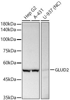
Western blot analysis of various lysates using GLUD2 Rabbit pAb (CAB6604) at 1:500 dilution. Secondary antibody: HRP-conjugated Goat anti-Rabbit IgG (H+L) (CABS014) at 1:10000 dilution. Lysates/proteins: 25μg per lane. Blocking buffer: 3% nonfat dry milk in TBST. Detection: ECL Basic Kit (AbGn00020). Exposure time: 10s.
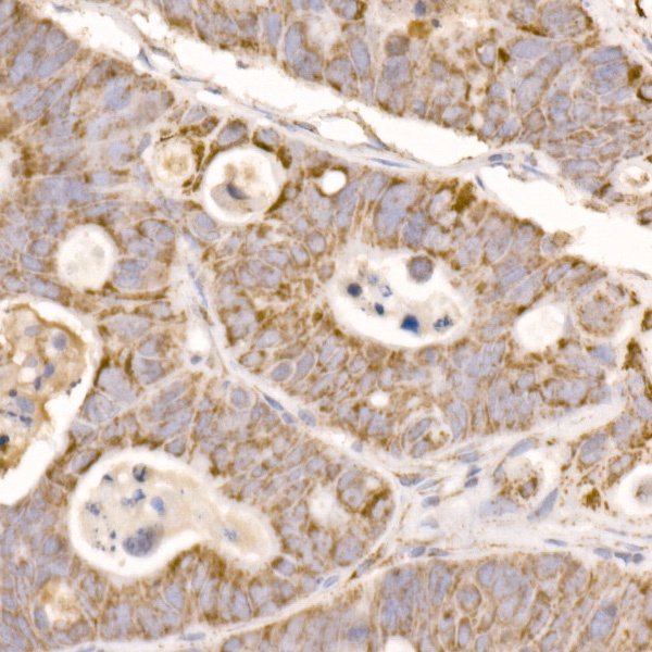
Immunohistochemistry analysis of paraffin-embedded Human colon carcinoma using GLUD2 Rabbit pAb (CAB6604) at dilution of 1:50 (40x lens). High pressure antigen retrieval performed with 0.01M Citrate buffer (pH 6.0) prior to IHC staining.
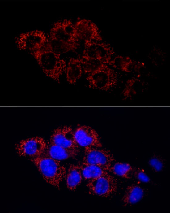
Immunofluorescence analysis of HepG2 cells using GLUD2 Rabbit pAb (CAB6604) at dilution of 1:50 (40x lens). Secondary antibody: Cy3-conjugated Goat anti-Rabbit IgG (H+L) (CABS007) at 1:500 dilution. Blue: DAPI for nuclear staining.
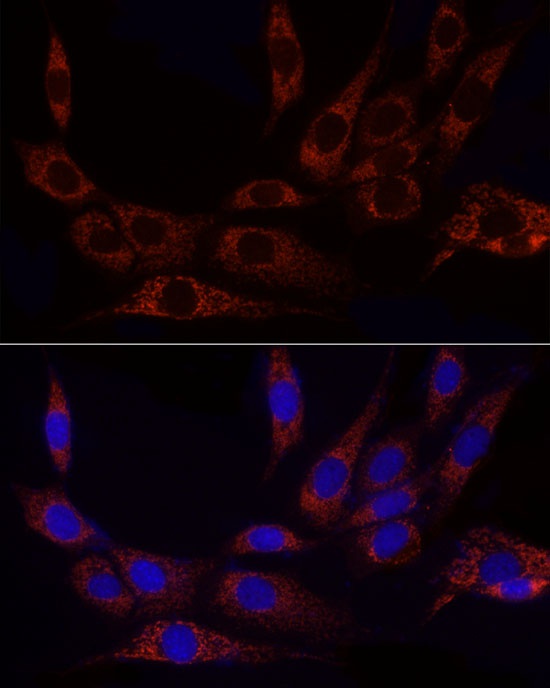
Immunofluorescence analysis of NIH/3T3 cells using GLUD2 Rabbit pAb (CAB6604) at dilution of 1:50 (40x lens). Secondary antibody: Cy3-conjugated Goat anti-Rabbit IgG (H+L) (CABS007) at 1:500 dilution. Blue: DAPI for nuclear staining.