The GP9 Antibody (CAB5374) is a high-quality antibody developed for reliable detection and analysis of target proteins. This antibody, produced in rabbits, exhibits high reactivity with human samples and has been validated for use in Western blot applications.By binding specifically to the GP9 protein, this antibody enables accurate detection and analysis of GP9 in various cell types, making it ideal for studies in hematology and cardiovascular research. Understanding the function of GP9 is essential for unraveling the mechanisms underlying platelet activation and thrombus formation, providing insights that could lead to the development of novel therapeutic strategies for various cardiovascular conditions.
This antibody is validated for use in WB, IHC-P, ELISA applications and has demonstrated reactivity against Human, Mouse, Rat samples.
Product Name:
GP9 Antibody
SKU:
CAB5374
Size:
20μL, 100μL
Reactivity:
Human, Mouse, Rat
Conjugate:
Unconjugated
Immunogen:
Recombinant protein (or fragment).This information is considered to be commercially sensitive.
Recommended starting concentration is 1 μg/mL. Please optimize the concentration based on your specific assay requirements.
Synonyms:
GPIX, CD42a, GP9
Positive Sample:
K-562
Cellular Localization:
Membrane, Single-Pass Type I Membrane Protein.
Calculated MW:
19kDa
Observed MW:
19kDa
This gene encodes a small membrane glycoprotein found on the surface of human platelets. It forms a 1-to-1 noncovalent complex with glycoprotein Ib, a platelet surface membrane glycoprotein complex that functions as a receptor for von Willebrand factor. The complete receptor complex includes noncovalent association of the alpha and beta subunits with the protein encoded by this gene and platelet glycoprotein V. Defects in this gene are a cause of Bernard-Soulier syndrome, also known as giant platelet disease. These patients have unusually large platelets and have a clinical bleeding tendency.
Purification Method
Affinity purification
Gene ID
2815
RRID
AB_2766184
Buffer Information
Store at -20℃. Avoid freeze / thaw cycles. Buffer: PBS containing 50% glycerol, preserved with proclin300 or sodium azide, pH 7.3.
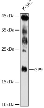
Western blot analysis of lysates from K-562 cells, using GP9 Rabbit pAb (CAB5374) at 1:500 dilution. Secondary antibody: HRP-conjugated Goat anti-Rabbit IgG (H+L) (CABS014) at 1:10000 dilution. Lysates/proteins: 25μg per lane. Blocking buffer: 3% nonfat dry milk in TBST. Detection: ECL Enhanced Kit (AbGn00021). Exposure time: 180s.
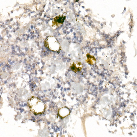
Immunohistochemistry analysis of paraffin-embedded Rat bone marrow using GP9 Rabbit pAb (CAB5374) at dilution of 1:20 (40x lens). High pressure antigen retrieval performed with 0.01M Citrate buffer (pH 6.0) prior to IHC staining.