The [KO Validated] macroH2A.1 Antibody (CAB7045) is a high-quality antibody developed for reliable detection and analysis of target proteins. This antibody is produced in rabbits and has been validated for use in Western blot applications, providing accurate and reliable results.H2AFY, also known as macroH2A, is a variant of the histone H2A protein that plays a crucial role in DNA packaging and gene expression regulation. Research has shown that H2AFY is involved in various cellular processes, including embryonic development, X chromosome inactivation, and tumor suppression.
This antibody is validated for use in WB, IHC-P, IF/ICC, ELISA applications and has demonstrated reactivity against Human, Mouse, Rat samples.
Product Name:
[KO Validated] macroH2A.1 Antibody
SKU:
CAB7045
Size:
20μL, 100μL
Reactivity:
Human, Mouse, Rat
Conjugate:
Unconjugated
Immunogen:
Recombinant protein (or fragment).This information is considered to be commercially sensitive.
Histones are basic nuclear proteins that are responsible for the nucleosome structure of the chromosomal fiber in eukaryotes. Nucleosomes consist of approximately 146 bp of DNA wrapped around a histone octamer composed of pairs of each of the four core histones (H2A, H2B, H3, and H4). The chromatin fiber is further compacted through the interaction of a linker histone, H1, with the DNA between the nucleosomes to form higher order chromatin structures. This gene encodes a replication-independent histone that is a member of the histone H2A family. It replaces conventional H2A histones in a subset of nucleosomes where it represses transcription and participates in stable X chromosome inactivation. Alternative splicing results in multiple transcript variants encoding different isoforms.
Purification Method
Affinity purification
Gene ID
9555
RRID
AB_2767600
Buffer Information
Store at -20℃. Avoid freeze / thaw cycles. Buffer: PBS containing 50% glycerol, preserved with proclin300 or sodium azide, pH 7.3.
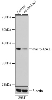
Western blot analysis of lysates from wild type (WT) and macroH2A.1 knockout (KO) 293T cells, using [KO Validated] macroH2A.1 Rabbit pAb (CAB7045) at 1:1000 dilution. Secondary antibody: HRP-conjugated Goat anti-Rabbit IgG (H+L) (CABS014) at 1:10000 dilution. Lysates/proteins: 25μg per lane. Blocking buffer: 3% nonfat dry milk in TBST. Detection: ECL Basic Kit (AbGn00020). Exposure time: 1s.
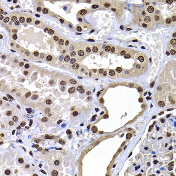
Immunohistochemistry analysis of paraffin-embedded Human kidney using [KO Validated] macroH2A.1 Rabbit pAb (CAB7045) at dilution of 1:100 (40x lens). Microwave antigen retrieval performed with 0.01M PBS Buffer (pH 7.2) prior to IHC staining.
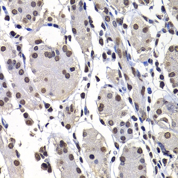
Immunohistochemistry analysis of paraffin-embedded Human stomach using [KO Validated] macroH2A.1 Rabbit pAb (CAB7045) at dilution of 1:100 (40x lens). Microwave antigen retrieval performed with 0.01M PBS Buffer (pH 7.2) prior to IHC staining.
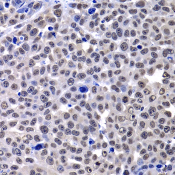
Immunohistochemistry analysis of paraffin-embedded Mouse lung cancer using [KO Validated] macroH2A.1 Rabbit pAb (CAB7045) at dilution of 1:100 (40x lens). Microwave antigen retrieval performed with 0.01M PBS Buffer (pH 7.2) prior to IHC staining.