The HADH Antibody (CAB1076) is a high-quality antibody developed for reliable detection and analysis of target proteins. This polyclonal antibody, raised in rabbits, is highly specific to human samples and has been validated for use in Western blot applications. By binding to the HADH protein, this antibody enables researchers to study its expression and activity in various cell types, making it ideal for investigations in metabolism, diabetes, and mitochondrial disorders.HADH, also known as hydroxyacyl-CoA dehydrogenase, is an enzyme involved in the breakdown of fatty acids for energy production.
This antibody is validated for use in WB, IHC-P, ELISA applications and has demonstrated reactivity against Human, Mouse, Rat samples.
Product Name:
HADH Antibody
SKU:
CAB1076
Size:
20μL, 100μL
Reactivity:
Human, Mouse, Rat
Conjugate:
Unconjugated
Immunogen:
Recombinant protein (or fragment).This information is considered to be commercially sensitive.
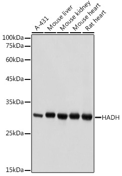
A-431, Mouse liver, Mouse kidney, Mouse heart, Rat heart
Cellular Localization:
Mitochondrion Matrix.
Calculated MW:
34kDa
Observed MW:
34kDa
This gene is a member of the 3-hydroxyacyl-CoA dehydrogenase gene family. The encoded protein functions in the mitochondrial matrix to catalyze the oxidation of straight-chain 3-hydroxyacyl-CoAs as part of the beta-oxidation pathway. Its enzymatic activity is highest with medium-chain-length fatty acids. Mutations in this gene cause one form of familial hyperinsulinemic hypoglycemia. The human genome contains a related pseudogene of this gene on chromosome 15.
Purification Method
Affinity purification
Gene ID
3033
RRID
AB_2758202
Buffer Information
Store at -20℃. Avoid freeze / thaw cycles. Buffer: PBS containing 50% glycerol, preserved with proclin300 or sodium azide, pH 7.3.
Western blot analysis of various lysates using HADH Rabbit pAb (CAB1076) at 1:1000 dilution. Secondary antibody: HRP-conjugated Goat anti-Rabbit IgG (H+L) (CABS014) at 1:10000 dilution. Lysates/proteins: 25μg per lane. Blocking buffer: 3% nonfat dry milk in TBST. Detection: ECL Basic Kit (AbGn00020). Exposure time: 1s.
Immunohistochemistry analysis of paraffin-embedded Rat spinal cord using HADH Rabbit pAb (CAB1076) at dilution of 1:100 (40x lens). Microwave antigen retrieval performed with 0.01M PBS Buffer (pH 7.2) prior to IHC staining.
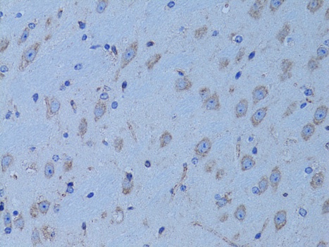
Immunohistochemistry analysis of paraffin-embedded Mouse brain using HADH Rabbit pAb (CAB1076) at dilution of 1:100 (40x lens). Microwave antigen retrieval performed with 0.01M PBS Buffer (pH 7.2) prior to IHC staining.