The KAT1/HAT1 Monoclonal Antibody (CAB4423) is a high-quality antibody developed for reliable detection and analysis of target proteins. This antibody, produced in rabbits, is highly specific to human samples and has been validated for use in Western blot applications. By binding to the KAT1 (HAT1) protein, this antibody allows for the accurate detection and analysis of KAT1 (HAT1) levels in various cell types, making it an invaluable resource for studies in epigenetics, chromatin remodeling, and gene expression regulation.
This antibody is validated for use in WB, IHC-P, IF/ICC, IP, ELISA applications and has demonstrated reactivity against Human, Mouse, Rat samples.
Product Name:
KAT1/HAT1 Monoclonal Antibody
SKU:
CAB4423
Size:
20μL, 100μL
Reactivity:
Human, Mouse, Rat
Clone Number:
ARC1002
Conjugate:
Unconjugated
Immunogen:
Recombinant protein (or fragment).This information is considered to be commercially sensitive.
0.5μg-4μg antibody for 200μg-400μg extracts of whole cells
IHC-P
1:50 - 1:200
IF/ICC
1:50 - 1:200
ELISA
Recommended starting concentration is 1 μg/mL. Please optimize the concentration based on your specific assay requirements.
Synonyms:
KAT1, KAT1/HAT1
Positive Sample:
MCF7, U-87MG, Mouse testis, Rat thymus, HeLa
Cellular Localization:
Cytoplasm, Nucleus, Nucleus Matrix, Nucleoplasm.
Calculated MW:
50kDa
Observed MW:
49kDa
The protein encoded by this gene is a type B histone acetyltransferase (HAT) that is involved in the rapid acetylation of newly synthesized cytoplasmic histones, which are in turn imported into the nucleus for de novo deposition onto nascent DNA chains. Histone acetylation, particularly of histone H4, plays an important role in replication-dependent chromatin assembly. Specifically, this HAT can acetylate soluble but not nucleosomal histone H4 at lysines 5 and 12, and to a lesser degree, histone H2A at lysine 5. Alternatively spliced transcript variants have been identified for this gene.
Purification Method
Affinity purification
Gene ID
8520
RRID
AB_2863271
Buffer Information
Store at -20℃. Avoid freeze / thaw cycles. Buffer: PBS containing 50% glycerol and 0.05% BSA, preserved with proclin300 or sodium azide, pH 7.3.
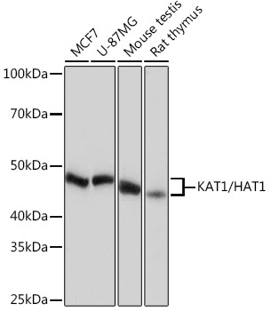
Western blot analysis of various lysates using [KO Validated] KAT1/HAT1 Rabbit mAb (CAB4423) at 1:1000 dilution. Secondary antibody: HRP-conjugated Goat anti-Rabbit IgG (H+L) (CABS014) at 1:10000 dilution. Lysates/proteins: 25 μg per lane. Blocking buffer: 3% nonfat dry milk in TBST. Detection: ECL Basic Kit (AbGn00020). Exposure time: 10 s.
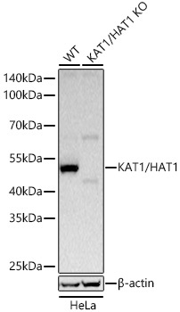
Western blot analysis of lysates from wild type (WT) and KAT1/HAT1 knockout (KO) HeLa cells using KAT1/HAT1 Rabbit mAb (CAB4423) at 1:1000 dilution incubated at room temperature for 1.5 hours. Secondary antibody: HRP-conjugated Goat anti-Rabbit IgG (H+L) (CABS014) at 1:10000 dilution. Lysates/proteins: 25 μg per lane. Blocking buffer: 3% nonfat dry milk in TBST. Detection: ECL Basic Kit (AbGn00020). Exposure time: 1 s.
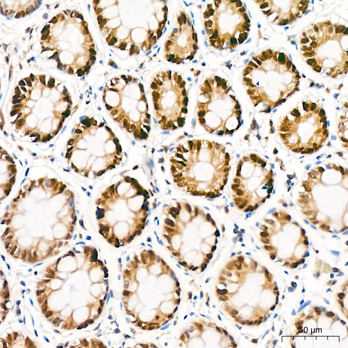
Immunohistochemistry analysis of paraffin-embedded Human colon tissue using [KO Validated] KAT1/HAT1 Rabbit mAb (CAB4423) at a dilution of 1:200 (40x lens). High pressure antigen retrieval performed with 0.01M Citrate buffer (pH 6.0) prior to IHC staining.
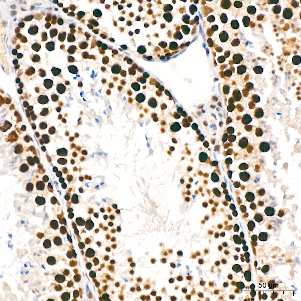
Immunohistochemistry analysis of paraffin-embedded Mouse testis tissue using [KO Validated] KAT1/HAT1 Rabbit mAb (CAB4423) at a dilution of 1:200 (40x lens). High pressure antigen retrieval performed with 0.01M Citrate buffer (pH 6.0) prior to IHC staining.
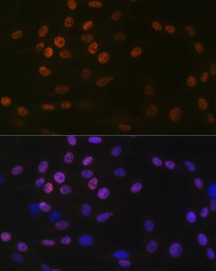
Immunofluorescence analysis of C6 cells using [KO Validated] KAT1/HAT1 Rabbit mAb (CAB4423) at dilution of 1:100 (40x lens). Secondary antibody: Cy3-conjugated Goat anti-Rabbit IgG (H+L) (CABS007) at 1:500 dilution. Blue: DAPI for nuclear staining.
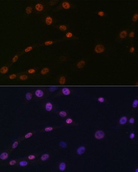
Immunofluorescence analysis of NIH/3T3 cells using [KO Validated] KAT1/HAT1 Rabbit mAb (CAB4423) at dilution of 1:100 (40x lens). Secondary antibody: Cy3-conjugated Goat anti-Rabbit IgG (H+L) (CABS007) at 1:500 dilution. Blue: DAPI for nuclear staining.