The [KO Validated] HDAC1 Antibody (CAB0238) is a high-quality antibody developed for reliable detection and analysis of target proteins. This antibody, produced in rabbits, is highly specific to human samples and has been validated for use in Western blot applications.HDAC1 is known to play a vital role in various biological processes, including cell cycle regulation, apoptosis, and differentiation. Dysregulation of HDAC1 has been implicated in several diseases, such as cancer, neurodegenerative disorders, and inflammatory conditions.
This antibody is validated for use in WB, IHC-P, IF/ICC, IP, ELISA applications and has demonstrated reactivity against Human, Mouse, Rat samples.
Product Name:
[KO Validated] HDAC1 Antibody
SKU:
CAB0238
Size:
20μL, 100μL
Reactivity:
Human, Mouse, Rat
Conjugate:
Unconjugated
Immunogen:
Synthetic peptide. This information is considered to be commercially sensitive.
0.5 μg - 4 μg antibody for 200 μg-400 μg extracts of whole cells
IF/ICC
1:50 - 1:200
IHC-P
1:50 - 1:200
ELISA
Recommended starting concentration is 1 μg/mL. Please optimize the concentration based on your specific assay requirements.
Synonyms:
HD1, RPD3, KDAC1, GON-10, RPD3L1, C1
Positive Sample:
293T, HeLa, NIH/3T3,HT-29
Cellular Localization:
Nucleus.
Calculated MW:
55kDa
Observed MW:
62kDa
Histone acetylation and deacetylation, catalyzed by multisubunit complexes, play a key role in the regulation of eukaryotic gene expression. The protein encoded by this gene belongs to the histone deacetylase/acuc/apha family and is a component of the histone deacetylase complex. It also interacts with retinoblastoma tumor-suppressor protein and this complex is a key element in the control of cell proliferation and differentiation. Together with metastasis-associated protein-2, it deacetylates p53 and modulates its effect on cell growth and apoptosis.
Purification Method
Affinity purification
Gene ID
3065
RRID
AB_2757051
Buffer Information
Store at -20℃. Avoid freeze / thaw cycles. Buffer: PBS containing 50% glycerol, preserved with proclin300 or sodium azide, pH 7.3.
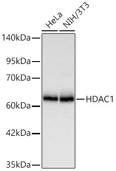
Western blot analysis of various lysates using [KD Validated] HDAC1 Rabbit pAb (CAB0238) at 1:2000 dilution incubated at room temperature for 1.5 hours. Secondary antibody: HRP-conjugated Goat anti-Rabbit IgG (H+L) (CABS014) at 1:10000 dilution. Lysates/proteins: 25 μg per lane. Blocking buffer: 3% nonfat dry milk in TBST. Detection: ECL Basic Kit (AbGn00020) Exposure time: 20 s.
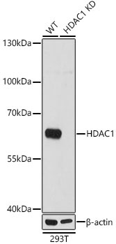
Western blot analysis of lysates from wild type (WT) and HDAC1 knockdown (KD) 293T cells, using [KD Validated] HDAC1 Rabbit pAb (CAB0238) at 1:1000 dilution. Secondary antibody: HRP-conjugated Goat anti-Rabbit IgG (H+L) (CABS014) at 1:10000 dilution. Lysates/proteins: 25μg per lane. Blocking buffer: 3% nonfat dry milk in TBST. Detection: ECL Basic Kit (AbGn00020). Exposure time: 1s.
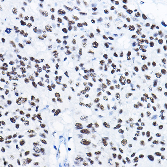
Immunohistochemistry analysis of paraffin-embedded Human lung cancer using [KD Validated] HDAC1 Rabbit pAb (CAB0238) at dilution of 1:50 (40x lens). High pressure antigen retrieval performed with 0.01M Citrate buffer (pH 6.0) prior to IHC staining.
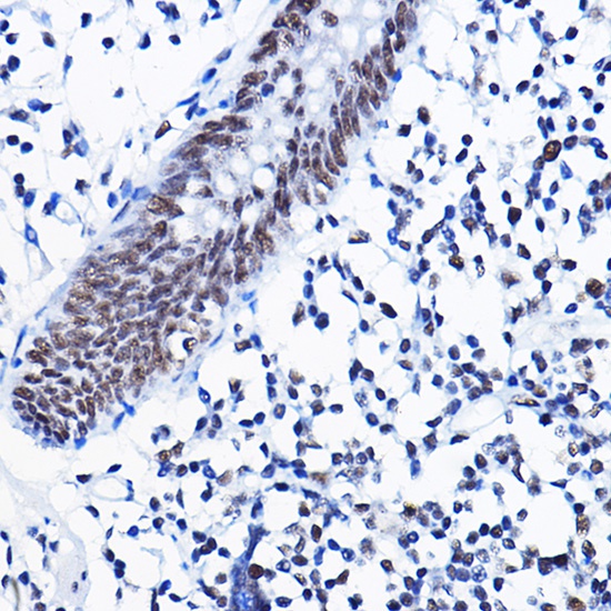
Immunohistochemistry analysis of paraffin-embedded Human colon using [KD Validated] HDAC1 Rabbit pAb (CAB0238) at dilution of 1:50 (40x lens). High pressure antigen retrieval performed with 0.01M Citrate buffer (pH 6.0) prior to IHC staining.
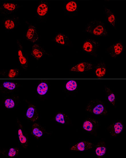
Confocal immunofluorescence analysis of L929 cells using [KD Validated] HDAC1 Rabbit pAb (CAB0238) at dilution of 1:200. Blue: DAPI for nuclear staining.
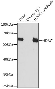
Immunoprecipitation analysis of 200 μg extracts of HT-29 cells, using 3 μg [KD Validated] HDAC1 Rabbit pAb (CAB0238). Western blot was performed from the immunoprecipitate using HDAC1 antibody (CAB0238) at a dilution of 1:1000.