The HDAC4 Antibody (CAB0179) is a high-quality antibody developed for reliable detection and analysis of target proteins. This antibody, raised in rabbits, is highly specific for human samples and has been validated for use in Western blot applications. It binds specifically to the HDAC4 protein, allowing for precise detection and analysis in a variety of cell types.HDAC4, a histone deacetylase, plays a crucial role in the regulation of gene expression and has been implicated in various diseases, including cancer and neurodegenerative disorders.
This antibody is validated for use in WB, IHC-P, ELISA applications and has demonstrated reactivity against Human, Mouse, Rat samples.
Product Name:
HDAC4 Antibody
SKU:
CAB0179
Size:
20μL, 100μL
Reactivity:
Human, Mouse, Rat
Conjugate:
Unconjugated
Immunogen:
Recombinant protein (or fragment).This information is considered to be commercially sensitive.
Histones play a critical role in transcriptional regulation, cell cycle progression, and developmental events. Histone acetylation/deacetylation alters chromosome structure and affects transcription factor access to DNA. The protein encoded by this gene belongs to class II of the histone deacetylase/acuc/apha family. It possesses histone deacetylase activity and represses transcription when tethered to a promoter. This protein does not bind DNA directly, but through transcription factors MEF2C and MEF2D. It seems to interact in a multiprotein complex with RbAp48 and HDAC3.
Purification Method
Affinity purification
Gene ID
9759
RRID
AB_2757003
Buffer Information
Store at -20℃. Avoid freeze / thaw cycles. Buffer: PBS containing 50% glycerol, preserved with proclin300 or sodium azide, pH 7.3.
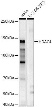
Western blot analysis of various lysates using HDAC4 Rabbit pAb (CAB0179) at 1:1000 dilution. Secondary antibody: HRP-conjugated Goat anti-Rabbit IgG (H+L) (CABS014) at 1:10000 dilution. Lysates/proteins: 25 μg per lane. Blocking buffer: 3% nonfat dry milk in TBST. Detection: ECL Basic Kit (AbGn00020). Negative control (NC): U-2 OS. Exposure time: 60s.
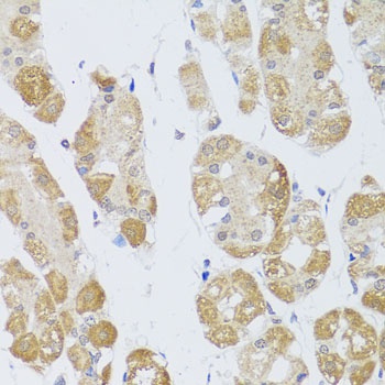
Immunohistochemistry analysis of paraffin-embedded Human stomach using HDAC4 Rabbit pAb (CAB0179) at dilution of 1:100 (40x lens). Microwave antigen retrieval performed with 0.01M PBS Buffer (pH 7.2) prior to IHC staining.
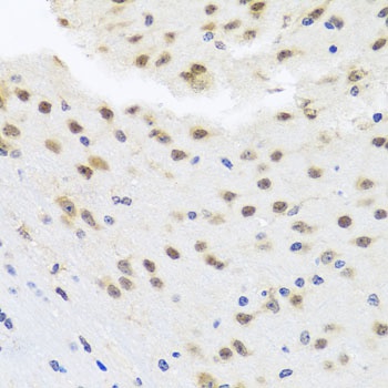
Immunohistochemistry analysis of paraffin-embedded Mouse brain using HDAC4 Rabbit pAb (CAB0179) at dilution of 1:100 (40x lens). Microwave antigen retrieval performed with 0.01M PBS Buffer (pH 7.2) prior to IHC staining.


")

