The HDAC8 Monoclonal Antibody (CAB8865) is a high-quality antibody developed for reliable detection and analysis of target proteins. This antibody, generated in rabbits, exhibits high specificity and sensitivity for detecting HDAC8 in human samples, making it an invaluable asset for experiments such as Western blot analysis.HDAC8 plays a crucial role in gene expression by removing acetyl groups from histone proteins, influencing chromatin structure and ultimately impacting cellular processes such as cell cycle regulation and development.
This antibody is validated for use in IHC-P, IF/ICC, IP, ELISA applications and has demonstrated reactivity against Human, Mouse, Rat samples.
Product Name:
HDAC8 Monoclonal Antibody
SKU:
CAB8865
Size:
20μL, 100μL
Reactivity:
Human, Mouse, Rat
Clone Number:
ARC1331
Conjugate:
Unconjugated
Immunogen:
Synthetic peptide. This information is considered to be commercially sensitive.
Histones play a critical role in transcriptional regulation, cell cycle progression, and developmental events. Histone acetylation/deacetylation alters chromosome structure and affects transcription factor access to DNA. The protein encoded by this gene belongs to class I of the histone deacetylase family. It catalyzes the deacetylation of lysine residues in the histone N-terminal tails and represses transcription in large multiprotein complexes with transcriptional co-repressors. Multiple transcript variants encoding different isoforms have been found for this gene.
Purification Method
Affinity purification
Gene ID
55869
RRID
AB_2863620
Buffer Information
Store at -20℃. Avoid freeze / thaw cycles. Buffer: PBS containing 50% glycerol and 0.05% BSA, preserved with proclin300 or sodium azide, pH 7.3.
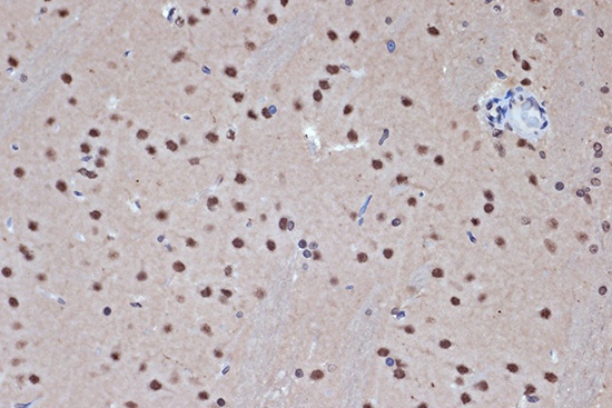
Immunohistochemistry analysis of paraffin-embedded Rat brain using HDAC8 Rabbit mAb (CAB8865) at dilution of 1:100 (40x lens). Microwave antigen retrieval performed with 0.01M Tris/EDTA Buffer (pH 9.0) prior to IHC staining.
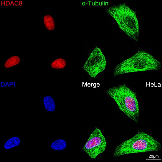
Confocal imaging of HeLa cells using HDAC8 Rabbit mAb (CAB8865, dilution 1:100) followed by a further incubation with Cy3-conjugated Goat Anti-Rabbit IgG (H+L) (CABS007, dilution 1:500) (Red). The cells were counterstained with α-Tubulin Mouse mAb (AC012, dilution 1:400) followed by incubation with ABflo® 488-conjugated Goat Anti-Mouse IgG (H+L) (CABS076, dilution 1:500) (Green). DAPI was used for nuclear staining (Blue). Objective: 100x.
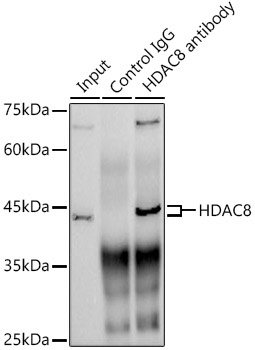
Immunoprecipitation analysis of 300 μg extracts from HeLa cells using 3 μg HDAC8 antibody (CAB8865). Western blot was performed from the immunoprecipitate using HDAC8 antibody (CAB8865) at a dilution of 1:1000.