The HIF1AN/FIH1 Polyclonal Antibody (CAB21823) is a high-quality antibody developed for reliable detection and analysis of target proteins. This protein enables 2-oxoglutarate-dependent dioxygenase activity and NF-κB binding, and is involved in the negative regulation of Notch signaling, transcriptional repression in response to hypoxia, and protein hydroxylation. It localizes to the cytosol, nucleoplasm, and perinuclear region, placing it at a key intersection of oxygen sensing, transcriptional control, and signaling pathway regulation.
This antibody is validated for use in WB, ELISA applications and has demonstrated reactivity against Human, Mouse samples.
Product Name:
HIF1AN/FIH1 Polyclonal Antibody
SKU:
CAB21823
Size:
20μL, 100μL
Reactivity:
Human, Mouse
Conjugate:
Unconjugated
Immunogen:
Recombinant protein (or fragment).This information is considered to be commercially sensitive.
Tested Applications:
WBELISA
Recommended Dilution:
WB
1:500 - 1:1000
ELISA
Recommended starting concentration is 1 μg/mL. Please optimize the concentration based on your specific assay requirements.
Synonyms:
FIH1, HIF1AN/FIH1
Positive Sample:
MCF7, Mouse heart, 3T3-L1
Cellular Localization:
Cytoplasm, Nucleus, Perinuclear Region.
Calculated MW:
41kDa
Observed MW:
40kDa
Enables several functions, including 2-oxoglutarate-dependent dioxygenase activity; NF-kappaB binding activity; and transition metal ion binding activity. Involved in several processes, including negative regulation of Notch signaling pathway; negative regulation of transcription from RNA polymerase II promoter in response to hypoxia; and protein hydroxylation. Located in cytosol; nucleoplasm; and perinuclear region of cytoplasm. Colocalizes with nucleus.
Purification Method
Affinity purification
Gene ID
55662
Buffer Information
Store at -20℃. Avoid freeze / thaw cycles. Buffer: PBS containing 50% glycerol, preserved with proclin300 or sodium azide, pH 7.3.
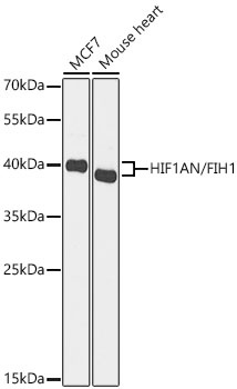
Western blot analysis of various lysates using HIF1AN/FIH1 Rabbit pAb (CAB21823) at 1:1000 dilution. Secondary antibody: HRP-conjugated Goat anti-Rabbit IgG (H+L) (CABS014) at 1:10000 dilution. Lysates / proteins: 25 μg per lane. Blocking buffer: 3 % nonfat dry milk in TBST. Detection: ECL Basic Kit (AbGn00020). Exposure time: 30s.
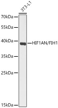
Western blot analysis of lysates from 3T3-L1 cells using HIF1AN/FIH1 Rabbit pAb (CAB21823) at 1:1000 dilution. Secondary antibody: HRP-conjugated Goat anti-Rabbit IgG (H+L) (CABS014) at 1:10000 dilution. Lysates/proteins: 25 μg per lane. Blocking buffer: 3% nonfat dry milk in TBST. Detection: ECL Basic Kit (AbGn00020). Exposure time: 90s.
")
")


")

