The [KO Validated] HIF1AN/FIH1 Antibody (CAB5466) is a high-quality antibody developed for reliable detection and analysis of target proteins. This antibody, produced in rabbits, is specifically designed for use in detecting and analyzing HIF1AN in human samples. Validated for Western blot applications, it provides researchers with a reliable means of studying the expression and function of HIF1AN in different cell types.HIF1AN, also known as FIH-1, acts as a negative regulator of the hypoxia inducible factor (HIF) pathway, influencing various cellular processes such as metabolism, angiogenesis, and apoptosis.
This antibody is validated for use in WB, IHC-P, IF/ICC, IP, ELISA applications and has demonstrated reactivity against Human, Mouse, Rat samples.
Product Name:
[KO Validated] HIF1AN/FIH1 Antibody
SKU:
CAB5466
Size:
20μL, 100μL
Reactivity:
Human, Mouse, Rat
Conjugate:
Unconjugated
Immunogen:
Recombinant protein (or fragment).This information is considered to be commercially sensitive.
Enables several functions, including 2-oxoglutarate-dependent dioxygenase activity; NF-kappaB binding activity; and transition metal ion binding activity. Involved in several processes, including negative regulation of Notch signaling pathway; negative regulation of transcription from RNA polymerase II promoter in response to hypoxia; and protein hydroxylation. Located in cytosol; nucleoplasm; and perinuclear region of cytoplasm. Colocalizes with nucleus.
Purification Method
Affinity purification
Gene ID
55662
RRID
AB_2766267
Buffer Information
Store at -20℃. Avoid freeze / thaw cycles. Buffer: PBS containing 50% glycerol, preserved with proclin300 or sodium azide, pH 7.3.
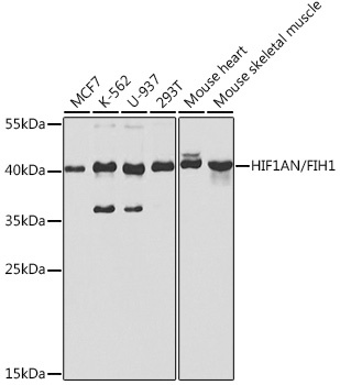
Western blot analysis of various lysates using [KO Validated] HIF1AN/FIH1 Rabbit pAb (CAB5466) at 1:1000 dilution. Secondary antibody: HRP-conjugated Goat anti-Rabbit IgG (H+L) (CABS014) at 1:10000 dilution. Lysates/proteins: 25μg per lane. Blocking buffer: 3% nonfat dry milk in TBST. Detection: ECL Basic Kit (AbGn00020). Exposure time: 30s.
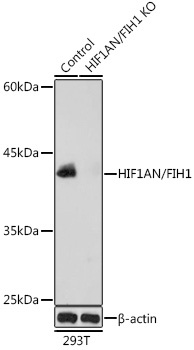
Western blot analysis of lysates from wild type (WT) and HIF1AN/FIH1 knockout (KO) 293T cells, using [KO Validated] HIF1AN/FIH1 Rabbit pAb (CAB5466) at 1:1000 dilution. Secondary antibody: HRP-conjugated Goat anti-Rabbit IgG (H+L) (CABS014) at 1:10000 dilution. Lysates/proteins: 25μg per lane. Blocking buffer: 3% nonfat dry milk in TBST. Detection: ECL Basic Kit (AbGn00020). Exposure time: 10s.
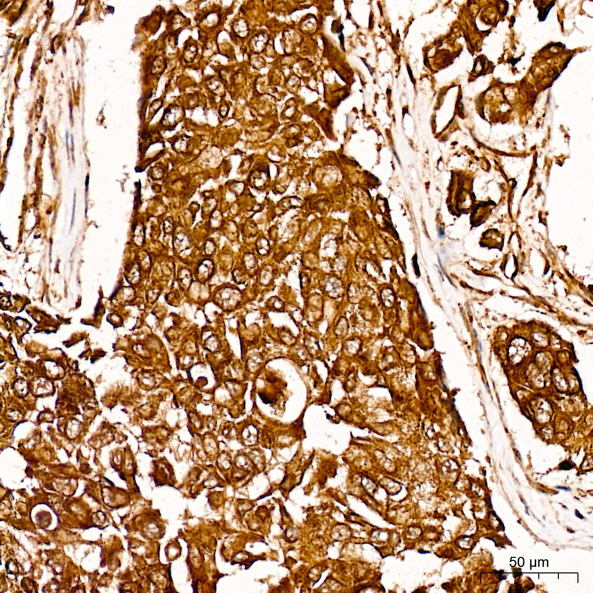
Immunohistochemistry analysis of paraffin-embedded Human breast cancer tissue using [KO Validated] HIF1AN/FIH1 Rabbit pAb (CAB5466) at a dilution of 1:100 (40x lens). High pressure antigen retrieval was performed with 0.01 M citrate buffer (pH 6.0) prior to IHC staining.
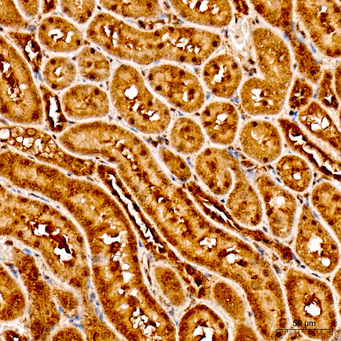
Immunohistochemistry analysis of paraffin-embedded Rat kidney tissue using [KO Validated] HIF1AN/FIH1 Rabbit pAb (CAB5466) at a dilution of 1:100 (40x lens). High pressure antigen retrieval was performed with 0.01 M citrate buffer (pH 6.0) prior to IHC staining.
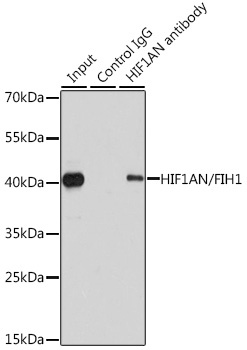
Immunoprecipitation analysis of 200 μg extracts of 293T cells using 1 μg HIF1AN/FIH1 antibody (CAB5466). Western blot was performed from the immunoprecipitate using HIF1AN/FIH1 antibody (CAB5466) at a dilution of 1:1000.


")


