The [KO Validated] HIF1AN/FIH1 Antibody (CAB5466) is a high-quality antibody developed for reliable detection and analysis of target proteins. Enables several functions, including 2-oxoglutarate-dependent dioxygenase activity; NF-kappaB binding activity; and transition metal ion binding activity. Involved in several processes, including negative regulation of Notch signaling pathway; negative regulation of transcription from RNA polymerase II promoter in response to hypoxia; and protein hydroxylation. Located in cytosol; nucleoplasm; and perinuclear region of cytoplasm. Colocalizes with nucleus.
This antibody is validated for use in WB, IHC-P, IF/ICC, IP, ELISA applications and has demonstrated reactivity against Human, Mouse, Rat samples.
Product Name:
[KO Validated] HIF1AN/FIH1 Antibody
SKU:
CAB5466
Size:
100μL, 20μL
Reactivity:
Human, Mouse, Rat
Conjugate:
Unconjugated
Immunogen:
Recombinant protein (or fragment).This information is considered to be commercially sensitive.
Tested Applications:
WBIHC-PIF/ICCIPELISA
Recommended Dilution:
WB
1:500 - 1:2000
IHC-P
1:50 - 1:200
IF/ICC
1:50 - 1:200
IP
0.5μg-4μg antibody for 200μg-400μg extracts of whole cells
ELISA
Recommended starting concentration is 1 μg/mL. Please optimize the concentration based on your specific assay requirements.
Enables several functions, including 2-oxoglutarate-dependent dioxygenase activity; NF-kappaB binding activity; and transition metal ion binding activity. Involved in several processes, including negative regulation of Notch signaling pathway; negative regulation of transcription from RNA polymerase II promoter in response to hypoxia; and protein hydroxylation. Located in cytosol; nucleoplasm; and perinuclear region of cytoplasm. Colocalizes with nucleus.
Purification Method
Affinity purification
Gene ID
55662
RRID
AB_2766267
Buffer Information
Store at -20℃. Avoid freeze / thaw cycles. Buffer: PBS containing 50% glycerol, preserved with proclin300 or sodium azide, pH 7.3.
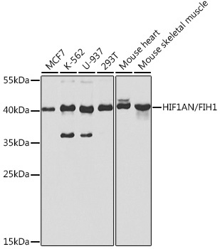
Western blot analysis of various lysates using [KO Validated] HIF1AN/FIH1 Rabbit pAb (CAB5466) at 1:1000 dilution. Secondary antibody: HRP-conjugated Goat anti-Rabbit IgG (H+L) (AS014) at 1:10000 dilution. Lysates/proteins: 25μg per lane. Blocking buffer: 3% nonfat dry milk in TBST. Detection: ECL Basic Kit (AbGn00020). Exposure time: 30s.
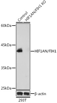
Western blot analysis of lysates from wild type (WT) and HIF1AN/FIH1 knockout (KO) 293T cells, using [KO Validated] HIF1AN/FIH1 Rabbit pAb (CAB5466) at 1:1000 dilution. Secondary antibody: HRP-conjugated Goat anti-Rabbit IgG (H+L) (AS014) at 1:10000 dilution. Lysates/proteins: 25μg per lane. Blocking buffer: 3% nonfat dry milk in TBST. Detection: ECL Basic Kit (AbGn00020). Exposure time: 10s.
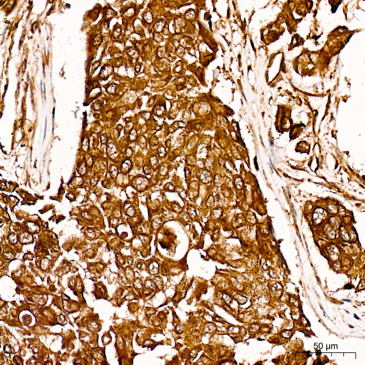
Immunohistochemistry analysis of paraffin-embedded Human breast cancer tissue using [KO Validated] HIF1AN/FIH1 Rabbit pAb (CAB5466) at a dilution of 1:100 (40x lens). High pressure antigen retrieval was performed with 0.01 M citrate buffer (pH 6.0) prior to IHC staining.
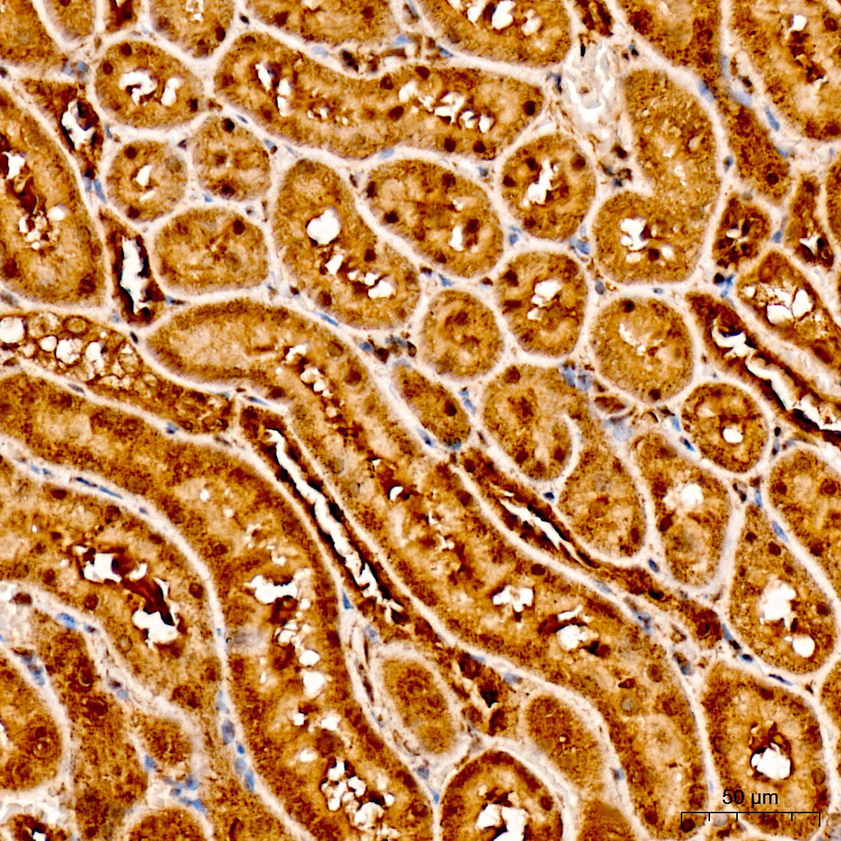
Immunohistochemistry analysis of paraffin-embedded Rat kidney tissue using [KO Validated] HIF1AN/FIH1 Rabbit pAb (CAB5466) at a dilution of 1:100 (40x lens). High pressure antigen retrieval was performed with 0.01 M citrate buffer (pH 6.0) prior to IHC staining.
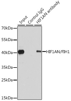
Immunoprecipitation analysis of 200 μg extracts of 293T cells using 1 μg HIF1AN/FIH1 antibody (CAB5466). Western blot was performed from the immunoprecipitate using HIF1AN/FIH1 antibody (CAB5466) at a dilution of 1:1000.


")


