The HIST1H3D Polyclonal Antibody (CAB21306) is a high-quality antibody developed for reliable detection and analysis of target proteins. This antibody, generated in rabbits, exhibits high specificity and sensitivity when used with human samples in Western blot applications.Hist1H3D is a histone protein that plays a crucial role in chromatin structure and gene regulation. It is involved in key cellular processes such as DNA replication, repair, and transcription. The Hist1H3D Polyclonal Antibody enables precise detection and analysis of Hist1H3D protein levels in various cell types, making it an essential tool for studies in epigenetics, gene expression, and cancer research.
This antibody is validated for use in WB, ELISA applications and has demonstrated reactivity against Human, Mouse, Rat, Other (Wide Range Predicted) samples.
Product Name:
HIST1H3D Polyclonal Antibody
SKU:
CAB21306
Size:
20μL, 100μL
Reactivity:
Human, Mouse, Rat, Other (Wide Range Predicted)
Immunogen:
Recombinant protein (or fragment).This information is considered to be commercially sensitive.
Histones are basic nuclear proteins that are responsible for the nucleosome structure of the chromosomal fiber in eukaryotes. Nucleosomes consist of approximately 146 bp of DNA wrapped around a histone octamer composed of pairs of each of the four core histones (H2A, H2B, H3, and H4). The chromatin fiber is further compacted through the interaction of a linker histone, H1, with the DNA between the nucleosomes to form higher order chromatin structures. This gene is intronless and encodes a replication-dependent histone that is a member of the histone H3 family. Transcripts from this gene lack polyA tails; instead, they contain a palindromic termination element. This gene is located separately from the other H3 genes that are in the histone gene cluster on chromosome 6p22-p21.3.
Purification Method
Affinity purification
Gene ID
8290 8350
Buffer Information
Store at -20℃. Avoid freeze / thaw cycles. Buffer: PBS containing 50% glycerol, preserved with proclin300 or sodium azide, pH 7.3.
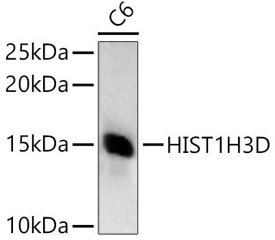
Western blot analysis of lysates from C6 cells, using HIST1H3D Rabbit pAb (CAB21306) at 1:1000 dilution. Secondary antibody: HRP-conjugated Goat anti-Rabbit IgG (H+L) (CABS014) at 1:10000 dilution. Lysates/proteins: 25μg per lane. Blocking buffer: 3% nonfat dry milk in TBST. Detection: ECL Basic Kit (AbGn00020). Exposure time: 5s.
 at 1:1000 dilution. Secondary antibody: HRP Goat Anti-Rabbit IgG (H+L) at 1:10000 dilution. Lysates/proteins: 25μg per lane. Blocking buffer: 3% nonfat dry milk in TBST.")
 at 1:1000 dilution. Secondary antibody: HRP Goat Anti-Rabbit IgG (H+L) at 1:10000 dilution. Lysates/proteins: 25μg per lane. Blocking buffer: 3% nonfat dry milk in TBST.")
