The [KO Validated] Histone H1.0 Antibody (CAB3298) is a high-quality antibody developed for reliable detection and analysis of target proteins. This polyclonal antibody, validated for knockout specificity, is specifically designed to target the Histone H1.0 protein and is highly reactive with human samples.Histone H1.0 is a variant of the histone H1 family that is involved in the packaging of DNA into nucleosomes and plays a critical role in the regulation of gene expression. By targeting Histone H1.0, researchers can investigate its role in chromatin organization and epigenetic regulation.
This antibody is validated for use in WB, IHC-P, ELISA applications and has demonstrated reactivity against Human, Mouse, Rat samples.
Product Name:
[KO Validated] Histone H1.0 Antibody
SKU:
CAB3298
Size:
20μL, 100μL
Reactivity:
Human, Mouse, Rat
Conjugate:
Unconjugated
Immunogen:
Recombinant protein (or fragment).This information is considered to be commercially sensitive.
Recommended starting concentration is 1 μg/mL. Please optimize the concentration based on your specific assay requirements.
Synonyms:
H10, H1.0, H1F0, H1FV, .0
Positive Sample:
293T
Cellular Localization:
Chromosome, Nucleus.
Calculated MW:
21kDa
Observed MW:
30kDa
Histones are basic nuclear proteins that are responsible for the nucleosome structure of the chromosomal fiber in eukaryotes. Nucleosomes consist of approximately 146 bp of DNA wrapped around a histone octamer composed of pairs of each of the four core histones (H2A, H2B, H3, and H4). The chromatin fiber is further compacted through the interaction of a linker histone, H1, with the DNA between the nucleosomes to form higher order chromatin structures. This gene is intronless and encodes a replication-independent histone that is a member of the histone H1 family.
Purification Method
Affinity purification
Gene ID
3005
RRID
AB_2765032
Buffer Information
Store at -20℃. Avoid freeze / thaw cycles. Buffer: PBS containing 50% glycerol, preserved with proclin300 or sodium azide, pH 7.3.
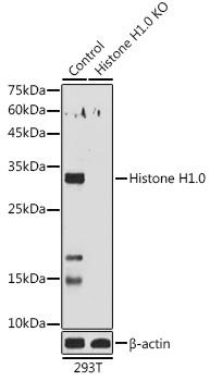
Western blot analysis of lysates from wild type (WT) and Histone H1.0 knockout (KO) 293T cells, using [KO Validated] Histone H1.0 Rabbit pAb (CAB3298) at 1:1000 dilution. Secondary antibody: HRP-conjugated Goat anti-Rabbit IgG (H+L) (CABS014) at 1:10000 dilution. Lysates/proteins: 25μg per lane. Blocking buffer: 3% nonfat dry milk in TBST. Detection: ECL Basic Kit (AbGn00020). Exposure time: 180s.
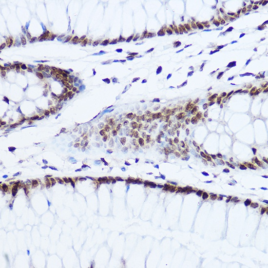
Immunohistochemistry analysis of paraffin-embedded Human colon using [KO Validated] Histone H1.0 Rabbit pAb (CAB3298) at dilution of 1:50 (40x lens). High pressure antigen retrieval performed with 0.01M Citrate buffer (pH 6.0) prior to IHC staining.
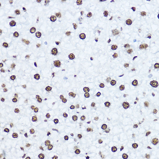
Immunohistochemistry analysis of paraffin-embedded Mouse liver using [KO Validated] Histone H1.0 Rabbit pAb (CAB3298) at dilution of 1:50 (40x lens). High pressure antigen retrieval performed with 0.01M Citrate buffer (pH 6.0) prior to IHC staining.
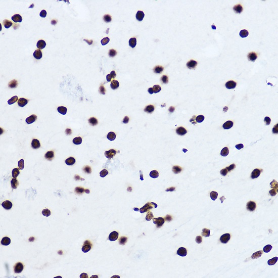
Immunohistochemistry analysis of paraffin-embedded Rat brain using [KO Validated] Histone H1.0 Rabbit pAb (CAB3298) at dilution of 1:50 (40x lens). High pressure antigen retrieval performed with 0.01M Citrate buffer (pH 6.0) prior to IHC staining.