The HLA-DPB1 Antibody (CAB17495) is a high-quality antibody developed for reliable detection and analysis of target proteins. This antibody, generated in rabbits, demonstrates high specificity and reactivity with human samples, making it a reliable tool for Western blot analysis.HLA-DPB1 plays a crucial role in presenting antigens to T cells, thereby modulating the adaptive immune response. Its polymorphisms have been linked to various autoimmune diseases and susceptibility to infections.
This antibody is validated for use in WB, IHC-P, IF/ICC, ELISA applications and has demonstrated reactivity against Human, Mouse, Rat samples.
Product Name:
HLA-DPB1 Antibody
SKU:
CAB17495
Size:
20μL, 100μL
Reactivity:
Human, Mouse, Rat
Conjugate:
Unconjugated
Immunogen:
Synthetic peptide. This information is considered to be commercially sensitive.
Recommended starting concentration is 1 μg/mL. Please optimize the concentration based on your specific assay requirements.
Synonyms:
DPB1, HLA-DP, HLA-DPB, HLA-DP1B, HLA-DPB1
Positive Sample:
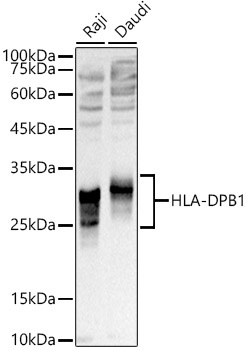
Raji, Daudi
Cellular Localization:
Cell Membrane, Endoplasmic Reticulum Membrane, Endosome Membrane, Golgi Apparatus, Lysosome Membrane, Single-Pass Type I Membrane Protein, Trans-Golgi Network Membrane.
Calculated MW:
29kDa
Observed MW:
25-35kDa
HLA-DPB belongs to the HLA class II beta chain paralogues. This class II molecule is a heterodimer consisting of an alpha (DPA) and a beta chain (DPB), both anchored in the membrane. It plays a central role in the immune system by presenting peptides derived from extracellular proteins. Class II molecules are expressed in antigen presenting cells (APC: B lymphocytes, dendritic cells, macrophages). The beta chain is approximately 26-28 kDa and its gene contains 6 exons. Exon one encodes the leader peptide, exons 2 and 3 encode the two extracellular domains, exon 4 encodes the transmembrane domain and exon 5 encodes the cytoplasmic tail. Within the DP molecule both the alpha chain and the beta chain contain the polymorphisms specifying the peptide binding specificities, resulting in up to 4 different molecules.
Purification Method
Affinity purification
Gene ID
3115
RRID
AB_2769801
Buffer Information
Store at -20℃. Avoid freeze / thaw cycles. Buffer: PBS containing 50% glycerol, preserved with proclin300 or sodium azide, pH 7.3.
Western blot analysis of various lysates using HLA-DPB1 Rabbit pAb (CAB17495) at 1:1000 dilution. Secondary antibody: HRP-conjugated Goat anti-Rabbit IgG (H+L) (CABS014) at 1:10000 dilution. Lysates/proteins: 25μg per lane. Blocking buffer: 3% nonfat dry milk in TBST. Detection: ECL Basic Kit (AbGn00020). Exposure time: 1s.
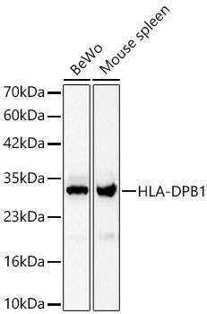
Western blot analysis of various lysates using HLA-DPB1 Rabbit pAb (CAB17495) at 1:1000 dilution. Secondary antibody: HRP-conjugated Goat anti-Rabbit IgG (H+L) (CABS014) at 1:10000 dilution. Lysates / proteins: 25 μg per lane. Blocking buffer: 3 % nonfat dry milk in TBST. Detection: ECL Basic Kit (AbGn00020). Exposure time: 45s.
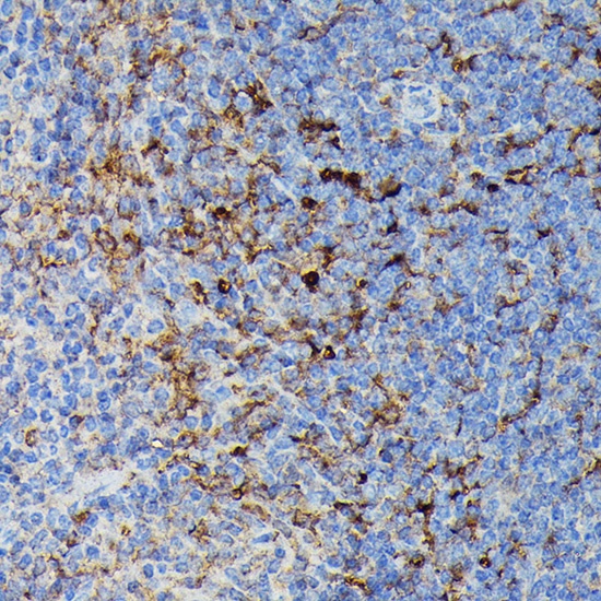
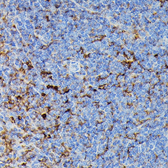
Immunohistochemistry analysis of paraffin-embedded Mouse spleen using HLA-DPB1 Rabbit pAb (CAB17495) at dilution of 1:100 (40x lens). High pressure antigen retrieval performed with 0.01M Citrate buffer (pH 6.0) prior to IHC staining.
Immunohistochemistry analysis of paraffin-embedded Rat spleen using HLA-DPB1 Rabbit pAb (CAB17495) at dilution of 1:100 (40x lens). High pressure antigen retrieval performed with 0.01M Citrate buffer (pH 6.0) prior to IHC staining.
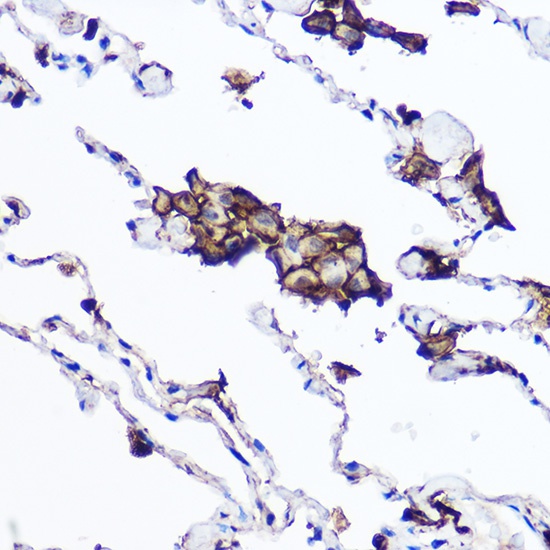
Immunohistochemistry analysis of paraffin-embedded Human lung tissue using HLA-DPB1 Rabbit pAb (CAB17495) at a dilution of 1:100 (40x lens). High pressure antigen retrieval was performed with 0.01 M citrate buffer (pH 6.0) prior to IHC staining.
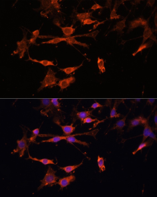
Immunofluorescence analysis of C6 cells using HLA-DPB1 Rabbit pAb (CAB17495) at dilution of 1:100. Secondary antibody: Cy3-conjugated Goat anti-Rabbit IgG (H+L) (CABS007) at 1:500 dilution. Blue: DAPI for nuclear staining.
Immunofluorescence analysis of HeLa cells using HLA-DPB1 Rabbit pAb (CAB17495) at dilution of 1:100. Secondary antibody: Cy3-conjugated Goat anti-Rabbit IgG (H+L) (CABS007) at 1:500 dilution. Blue: DAPI for nuclear staining.
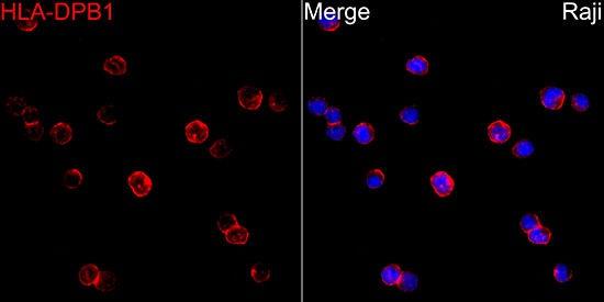
Immunofluorescence analysis of Raji cells using HLA-DPB1 Rabbit pAb (CAB17495) at dilution of 1:100 (40x lens). Secondary antibody: Cy3-conjugated Goat anti-Rabbit IgG (H+L) (CABS007) at 1:500 dilution. Blue: DAPI for nuclear staining.