The HNF4G Antibody (CAB16051) is a high-quality antibody developed for reliable detection and analysis of target proteins. This antibody, generated in rabbits, exhibits high reactivity with human samples and is validated for use in Western blot applications. By targeting the HNF4G protein, researchers can detect and analyze its expression in different cell types, making it invaluable for studies in molecular biology and metabolic research.HNF4G, a member of the hepatocyte nuclear factor family, plays a critical role in controlling the expression of genes involved in glucose and lipid metabolism, as well as cell differentiation and proliferation.
This antibody is validated for use in WB, IF/ICC, ELISA applications and has demonstrated reactivity against Human, Mouse, Rat samples.
Product Name:
HNF4G Antibody
SKU:
CAB16051
Size:
20μL, 100μL
Reactivity:
Human, Mouse, Rat
Conjugate:
Unconjugated
Immunogen:
Recombinant protein (or fragment).This information is considered to be commercially sensitive.
Sequence:
ASND GSHL HHPM HPHL SQDP LTGQ TILL GPMS TLVH ADQI STPE TPLP SPPQ GSGQ EQYK IAAN QASV ISHQ HLSK QKQL
Tested Applications:
WBIF/ICCELISA
Recommended Dilution:
WB
1:1000 - 1:5000
IF/ICC
1:50 - 1:100
ELISA
Recommended starting concentration is 1 μg/mL. Please optimize the concentration based on your specific assay requirements.
Synonyms:
NR2A2, NR2A3, HNF4G
Positive Sample:
Mouse large intestine, Rat liver
Cellular Localization:
Nucleus.
Calculated MW:
46kDa
Observed MW:
46kDa/50kDa
Enables DNA-binding transcription activator activity, RNA polymerase II-specific and RNA polymerase II cis-regulatory region sequence-specific DNA binding activity. Involved in positive regulation of transcription by RNA polymerase II. Located in several cellular components, including intercellular bridge; mitotic spindle; and nucleoplasm.
Purification Method
Affinity purification
Gene ID
3174
RRID
AB_2763490
Buffer Information
Store at -20℃. Avoid freeze / thaw cycles. Buffer: PBS containing 50% glycerol, preserved with proclin300 or sodium azide, pH 7.3.
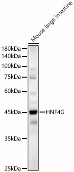
Western blot analysis of lysates from Mouse large intestine, using HNF4G Rabbit pAb (CAB16051) at 1:2000 dilution. Secondary antibody: HRP-conjugated Goat anti-Rabbit IgG (H+L) (CABS014) at 1:10000 dilution. Lysates/proteins: 25μg per lane. Blocking buffer: 3% nonfat dry milk in TBST. Detection: ECL Basic Kit (AbGn00020). Exposure time: 180s.
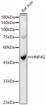
Western blot analysis of lysates from Rat liver, using HNF4G Rabbit pAb (CAB16051) at 1:2000 dilution. Secondary antibody: HRP-conjugated Goat anti-Rabbit IgG (H+L) (CABS014) at 1:10000 dilution. Lysates/proteins: 25μg per lane. Blocking buffer: 3% nonfat dry milk in TBST. Detection: ECL Basic Kit (AbGn00020). Exposure time: 60s.