The hnRNP A1 Antibody (CAB7491) is a high-quality antibody developed for reliable detection and analysis of target proteins. This antibody, produced in rabbits, exhibits high reactivity with human samples and is suitable for use in Western blot applications. By specifically binding to the HNRNPA1 protein, this antibody enables the detection and analysis of HNRNPA1 in various cell types, making it an ideal choice for investigations in molecular biology and cancer research.
This antibody is validated for use in WB, IHC-P, IF/ICC, ELISA applications and has demonstrated reactivity against Human, Mouse, Rat samples.
Product Name:
hnRNP A1 Antibody
SKU:
CAB7491
Size:
20μL, 100μL
Reactivity:
Human, Mouse, Rat
Conjugate:
Unconjugated
Immunogen:
Recombinant protein (or fragment).This information is considered to be commercially sensitive.
Recommended starting concentration is 1 μg/mL. Please optimize the concentration based on your specific assay requirements.
Synonyms:
UP 1, ALS19, ALS20, HNRPA1, IBMPFD3, HNRPA1L3, hnRNP A1, hnRNP-A1
Positive Sample:
A-549, Jurkat, HepG2, HL-60, 293T
Cellular Localization:
Cytoplasm, Nucleus.
Calculated MW:
39kDa
Observed MW:
39kDa
This gene encodes a member of a family of ubiquitously expressed heterogeneous nuclear ribonucleoproteins (hnRNPs), which are RNA-binding proteins that associate with pre-mRNAs in the nucleus and influence pre-mRNA processing, as well as other aspects of mRNA metabolism and transport. The protein encoded by this gene is one of the most abundant core proteins of hnRNP complexes and plays a key role in the regulation of alternative splicing. Mutations in this gene have been observed in individuals with amyotrophic lateral sclerosis 20. Multiple alternatively spliced transcript variants have been found. There are numerous pseudogenes of this gene distributed throughout the genome.
Purification Method
Affinity purification
Gene ID
3178
RRID
AB_2768022
Buffer Information
Store at -20℃. Avoid freeze / thaw cycles. Buffer: PBS containing 50% glycerol, preserved with proclin300 or sodium azide, pH 7.3.
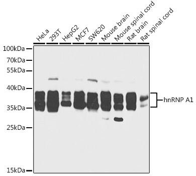
Western blot analysis of various lysates using hnRNP A1 Rabbit pAb (CAB7491) at 1:1000 dilution. Secondary antibody: HRP-conjugated Goat anti-Rabbit IgG (H+L) (CABS014) at 1:10000 dilution. Lysates/proteins: 25μg per lane. Blocking buffer: 3% nonfat dry milk in TBST. Detection: ECL Basic Kit (AbGn00020). Exposure time: 30s.
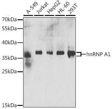
Western blot analysis of various lysates using hnRNP A1 Rabbit pAb (CAB7491) at 1:200 dilution. Secondary antibody: HRP-conjugated Goat anti-Rabbit IgG (H+L) (CABS014) at 1:10000 dilution. Lysates/proteins: 25μg per lane. Blocking buffer: 3% nonfat dry milk in TBST. Detection: ECL Basic Kit (AbGn00020). Exposure time: 5min.
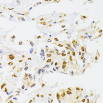
Immunohistochemistry analysis of paraffin-embedded Human breast cancer using hnRNP A1 Rabbit pAb (CAB7491) at dilution of 1:100 (40x lens). Microwave antigen retrieval performed with 0.01M PBS Buffer (pH 7.2) prior to IHC staining.
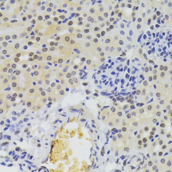
Immunohistochemistry analysis of paraffin-embedded Mouse kidney using hnRNP A1 Rabbit pAb (CAB7491) at dilution of 1:100 (40x lens). Microwave antigen retrieval performed with 0.01M PBS Buffer (pH 7.2) prior to IHC staining.
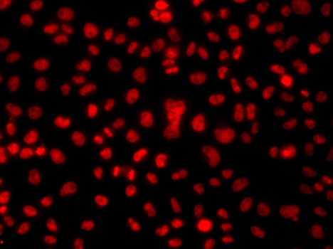
Immunofluorescence analysis of A549 cells using hnRNP A1 Rabbit pAb (CAB7491).Secondary antibody: Cy3-conjugated Goat anti-Rabbit IgG (H+L) (CABS007) at 1:500 dilution.