The HSCB Antibody (CAB15961) is a high-quality antibody developed for reliable detection and analysis of target proteins. This antibody, raised in rabbits, has been validated for use in various applications, including Western blot and immunohistochemistry.HSCB, also known as HscB chaperone protein, is essential for the proper assembly of iron-sulfur clusters in proteins involved in electron transport and oxidative phosphorylation. Dysfunction of HSCB can lead to mitochondrial disorders and metabolic diseases. Therefore, studying the role of HSCB is important for understanding mitochondrial function and developing potential treatments for related diseases.
This antibody is validated for use in WB, ELISA applications and has demonstrated reactivity against Human samples.
Product Name:
HSCB Antibody
SKU:
CAB15961
Size:
20μL, 100μL
Reactivity:
Human
Conjugate:
Unconjugated
Immunogen:
Recombinant protein (or fragment).This information is considered to be commercially sensitive.
Recommended starting concentration is 1 μg/mL. Please optimize the concentration based on your specific assay requirements.
Synonyms:
JAC1, HSC20, SIDBA5, DNAJC20, HSCB
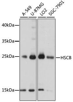
Positive Sample:
A-549, U-87MG, LO2, SGC-7901
Cellular Localization:
Cytoplasm, Mitochondrion.
Calculated MW:
27kDa
Observed MW:
27kDa
This gene encodes a DnaJ-type co-chaperone and member of the heat shock cognate B (HscB) family of proteins. The encoded protein plays a role in the synthesis of iron-sulfur clusters, protein cofactors that are involved in the redox reactions of mitochondrial electron transport and other processes. Cells in which this gene is knocked down exhibit reduced activity of iron-sulfur cluster-dependent enzymes including succinate dehydrogenase and aconitase. The encoded protein may stimulate the ATPase activity of the mitochondrial stress-70 protein. Alternative splicing results in multiple transcript variants.
Purification Method
Affinity purification
Gene ID
150274
RRID
AB_2763398
Buffer Information
Store at -20℃. Avoid freeze / thaw cycles. Buffer: PBS with 0.01% thimerosal,50% glycerol,pH7.3.
Western blot analysis of various lysates using HSCB Rabbit pAb (CAB15961) at 1000 dilution. Secondary antibody: HRP-conjugated Goat anti-Rabbit IgG (H+L) (CABS014) at 1:10000 dilution. Lysates/proteins: 25μg per lane. Blocking buffer: 3% nonfat dry milk in TBST. Detection: ECL Basic Kit (AbGn00020). Exposure time: 5s.