The IK Antibody (CAB15280) is a high-quality antibody developed for reliable detection and analysis of target proteins. This rabbit-derived antibody is highly specific to human samples and has been validated for use in Western blot applications. By binding to the IK protein, this antibody allows for the detection and analysis of IK in various cell types, making it ideal for studies in immunology and disease research.IK, also known as immune kinase, is essential for regulating the activity of immune cells and controlling the production of inflammatory molecules. Dysregulation of IK has been implicated in a variety of diseases, including autoimmune disorders, inflammatory conditions, and cancer.
This antibody is validated for use in WB, ELISA applications and has demonstrated reactivity against Human, Mouse, Rat samples.
Product Name:
IK Antibody
SKU:
CAB15280
Size:
20μL, 100μL
Reactivity:
Human, Mouse, Rat
Conjugate:
Unconjugated
Immunogen:
Recombinant protein (or fragment).This information is considered to be commercially sensitive.
Recommended starting concentration is 1 μg/mL. Please optimize the concentration based on your specific assay requirements.
Synonyms:
RED, RER, CSA2, IK
Positive Sample:
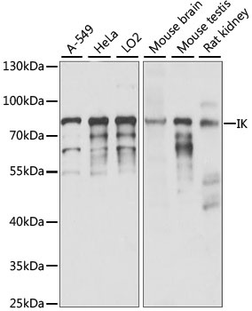
A-549, HeLa, LO2, Mouse brain, Mouse testis, Rat kidney
Cellular Localization:
Nucleus.
Calculated MW:
66kDa
Observed MW:
75kDa
The protein encoded by this gene was identified by its RED repeat, a stretch of repeated arginine, glutamic acid and aspartic acid residues. The protein localizes to discrete dots within the nucleus, excluding the nucleolus. Its function is unknown. This gene maps to chromosome 5; however, a pseudogene may exist on chromosome 2.
Purification Method
Affinity purification
Gene ID
3550
RRID
AB_2762180
Buffer Information
Store at -20℃. Avoid freeze / thaw cycles. Buffer: PBS with 0.01% thimerosal,50% glycerol,pH7.3.
Western blot analysis of various lysates using IK Rabbit pAb (CAB15280) at 1:1000 dilution. Secondary antibody: HRP-conjugated Goat anti-Rabbit IgG (H+L) (CABS014) at 1:10000 dilution. Lysates/proteins: 25μg per lane. Blocking buffer: 3% nonfat dry milk in TBST. Detection: ECL Enhanced Kit (AbGn00021). Exposure time: 15S.