The IL1RL1 Antibody (CAB1913) is a high-quality antibody developed for reliable detection and analysis of target proteins. IL1RL1 is known to play a crucial role in inflammatory responses and immune regulation. This antibody, generated in rabbits, is highly specific to human IL1RL1 and has been validated for use in applications such as Western blotting. By binding to the IL1RL1 protein, this antibody enables the detection and analysis of IL1RL1 expression in various cell types, making it an invaluable tool for studies in immunology, inflammation, and related fields.
This antibody is validated for use in WB, ELISA applications and has demonstrated reactivity against Human, Mouse, Rat samples.
Product Name:
IL1RL1 Antibody
SKU:
CAB1913
Size:
20μL, 100μL
Reactivity:
Human, Mouse, Rat
Conjugate:
Unconjugated
Immunogen:
Recombinant protein (or fragment).This information is considered to be commercially sensitive.
Recommended starting concentration is 1 μg/mL. Please optimize the concentration based on your specific assay requirements.
Synonyms:
T1, ST2, DER4, ST2L, ST2V, FIT-1, IL33R, IL1RL1
Positive Sample:
Mouse brain, HeLa, Hep G2, Mouse liver
Cellular Localization:
Cell Membrane, Secreted, Single-Pass Type I Membrane Protein.
Calculated MW:
63kDa
Observed MW:
30kDa/41kDa/70kDa
The protein encoded by this gene is a member of the interleukin 1 receptor family. Studies of the similar gene in mouse suggested that this receptor can be induced by proinflammatory stimuli, and may be involved in the function of helper T cells. This gene, interleukin 1 receptor, type I (IL1R1), interleukin 1 receptor, type II (IL1R2) and interleukin 1 receptor-like 2 (IL1RL2) form a cytokine receptor gene cluster in a region mapped to chromosome 2q12. Alternative splicing of this gene results in multiple transcript variants.
Purification Method
Affinity purification
Gene ID
9173
RRID
AB_2763941
Buffer Information
Store at -20℃. Avoid freeze / thaw cycles. Buffer: PBS containing 50% glycerol, preserved with proclin300 or sodium azide, pH 7.3.
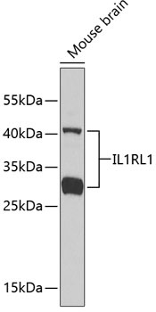
Western blot analysis of lysates from mouse brain, using IL1RL1 Rabbit pAb (CAB1913). Secondary antibody: HRP-conjugated Goat anti-Rabbit IgG (H+L) (CABS014) at 1:10000 dilution. Lysates/proteins: 25μg per lane. Blocking buffer: 3% nonfat dry milk in TBST.
Western blot analysis of various lysates using IL1RL1 Rabbit pAb (CAB1913) at 1:1000 dilution. Secondary antibody: HRP-conjugated Goat anti-Rabbit IgG (H+L) (CABS014) at 1:10000 dilution. Lysates / proteins: 25 μg per lane. Blocking buffer: 3 % nonfat dry milk in TBST. Detection: ECL Basic Kit (AbGn00020). Exposure time: 60s.