The [KO Validated] ING5 Antibody (CAB7288) is a high-quality antibody developed for reliable detection and analysis of target proteins. This antibody, generated in rabbits, has high reactivity with human samples and has been validated for use in Western blot applications. By binding to the ING5 protein, this antibody enables accurate detection and analysis in various cell types, making it an ideal tool for studies in cancer research and cell biology.ING5 is known to interact with histone deacetylases and histone acetyltransferases, playing a key role in chromatin remodeling and gene transcription regulation.
This antibody is validated for use in WB, IF/ICC, ELISA applications and has demonstrated reactivity against Human samples.
Product Name:
[KO Validated] ING5 Antibody
SKU:
CAB7288
Size:
20μL, 100μL
Reactivity:
Human
Conjugate:
Unconjugated
Immunogen:
Recombinant protein (or fragment).This information is considered to be commercially sensitive.
Recommended starting concentration is 1 μg/mL. Please optimize the concentration based on your specific assay requirements.
Synonyms:
p28ING5, G5
Positive Sample:
293T
Cellular Localization:
Nucleus.
Calculated MW:
28kDa
Observed MW:
30kDa
This gene encodes a tumor suppressor protein that inhibits cell growth and induces apoptosis. This protein contains a PHD-type zinc finger. It interacts with tumor suppressor p53 and p300, a component of the histone acetyl transferase complex, suggesting a role in transcriptional regulation. Alternative splicing and the use of multiple promoters and 3' terminal exons results in multiple transcript variants.
Purification Method
Affinity purification
Gene ID
84289
RRID
AB_2767829
Buffer Information
Store at -20℃. Avoid freeze / thaw cycles. Buffer: PBS containing 50% glycerol, preserved with proclin300 or sodium azide, pH 7.3.
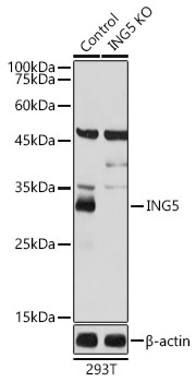
Western blot analysis of lysates from wild type (WT) and ING5 knockout (KO) 293T cells, using [KO Validated] ING5 Rabbit pAb (CAB7288) at 1:1000 dilution. Secondary antibody: HRP-conjugated Goat anti-Rabbit IgG (H+L) (CABS014) at 1:10000 dilution. Lysates/proteins: 25μg per lane. Blocking buffer: 3% nonfat dry milk in TBST. Detection: ECL Basic Kit (AbGn00020). Exposure time: 1s.
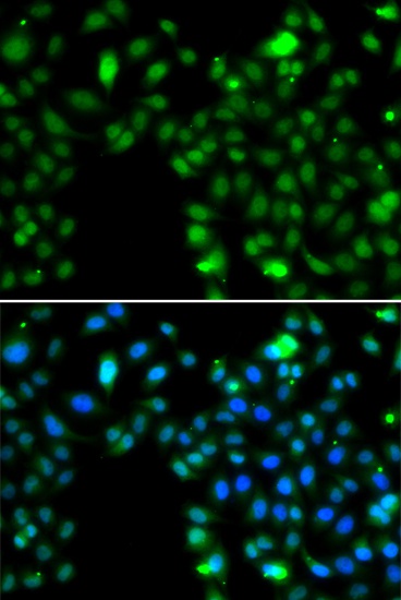
Immunofluorescence analysis of MCF-7 cells using [KO Validated] ING5 Rabbit pAb (CAB7288). Secondary antibody: Cy3-conjugated Goat anti-Rabbit IgG (H+L) (CABS007) at 1:500 dilution. Blue: DAPI for nuclear staining.