The INPP5J Antibody (CAB6631) is a high-quality antibody developed for reliable detection and analysis of target proteins. Predicted to enable phosphatidylinositol-3,4,5-trisphosphate 5-phosphatase activity and phosphatidylinositol-4,5-bisphosphate 5-phosphatase activity. Predicted to be involved in inositol phosphate dephosphorylation; negative regulation of peptidyl-serine phosphorylation; and phosphatidylinositol dephosphorylation. Predicted to act upstream of or within negative regulation of neuron projection development. Located in cytoplasm and ruffle.
This antibody is validated for use in WB, IF/ICC, ELISA applications and has demonstrated reactivity against Human, Mouse, Rat samples.
Product Name:
INPP5J Antibody
SKU:
CAB6631
Size:
100μL, 20μL
Reactivity:
Human, Mouse, Rat
Conjugate:
Unconjugated
Immunogen:
Recombinant protein (or fragment).This information is considered to be commercially sensitive.
Tested Applications:
WBIF/ICCELISA
Recommended Dilution:
WB
1:500 - 1:2000
IF/ICC
1:10 - 1:100
ELISA
Recommended starting concentration is 1 μg/mL. Please optimize the concentration based on your specific assay requirements.
Synonyms:
PIPP, INPP5, PIB5PA, INPP5J
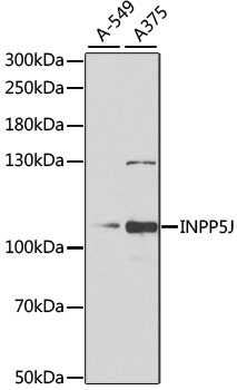
Positive Sample:
A-549, A375
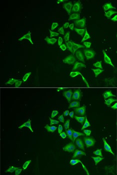
Cellular Localization:
Cytoplasm.
Calculated MW:
107kDa
Observed MW:
107kDa
Predicted to enable phosphatidylinositol-3,4,5-trisphosphate 5-phosphatase activity and phosphatidylinositol-4,5-bisphosphate 5-phosphatase activity. Predicted to be involved in inositol phosphate dephosphorylation; negative regulation of peptidyl-serine phosphorylation; and phosphatidylinositol dephosphorylation. Predicted to act upstream of or within negative regulation of neuron projection development. Located in cytoplasm and ruffle.
Purification Method
Affinity purification
Gene ID
27124
RRID
AB_2767220
Buffer Information
Store at -20℃. Avoid freeze / thaw cycles. Buffer: PBS containing 50% glycerol, preserved with proclin300 or sodium azide, pH 7.3.
Western blot analysis of various lysates using INPP5J Rabbit pAb (CAB6631) at 1:1000 dilution. Secondary antibody: HRP-conjugated Goat anti-Rabbit IgG (H+L) (AS014) at 1:10000 dilution. Lysates/proteins: 25μg per lane. Blocking buffer: 3% nonfat dry milk in TBST. Detection: ECL Basic Kit (AbGn00020). Exposure time: 90s.
Immunofluorescence analysis of U2OS cells using INPP5J Rabbit pAb (CAB6631). Secondary antibody: Cy3-conjugated Goat anti-Rabbit IgG (H+L) (AS007) at 1:500 dilution. Blue: DAPI for nuclear staining.