The [KD Validated] NUR77 Monoclonal Antibody (CAB24016) is a high-quality antibody developed for reliable detection and analysis of target proteins. This antibody, developed through a rigorous validation process, specifically recognizes Nur77 in human samples and is suitable for use in Western blot applications.Nur77, also known as NR4A1, is a key regulator of gene expression and cell signaling pathways that play crucial roles in various physiological and pathological conditions. By targeting Nur77 with this monoclonal antibody, researchers can gain valuable insights into the function and regulation of this important protein, making it an essential tool for studies in cell biology, cancer research, and drug discovery.
This antibody is validated for use in WB, ELISA applications and has demonstrated reactivity against Human, Mouse samples.
Product Name:
[KD Validated] NUR77 Monoclonal Antibody
SKU:
CAB24016
Size:
20μL, 100μL
Reactivity:
Human, Mouse
Clone Number:
ARC58221
Conjugate:
Unconjugated
Immunogen:
Synthetic peptide. This information is considered to be commercially sensitive.
This gene encodes a member of the steroid-thyroid hormone-retinoid receptor superfamily. Expression is induced by phytohemagglutinin in human lymphocytes and by serum stimulation of arrested fibroblasts. The encoded protein acts as a nuclear transcription factor. Translocation of the protein from the nucleus to mitochondria induces apoptosis. Multiple transcript variants encoding different isoforms have been found for this gene.
Purification Method
Affinity purification
Gene ID
3164
Buffer Information
Store at -20℃. Avoid freeze / thaw cycles. Buffer: PBS with 0.09% Sodium azide,0.05% BSA,50% glycerol,pH7.3.
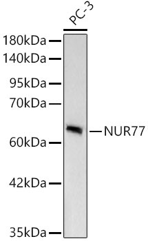
Western blot analysis of lysates from PC-3 cells, using [KD Validated] NUR77 Rabbit mAb (CAB24016) at 1:1000 dilution. Secondary antibody: HRP-conjugated Goat anti-Rabbit IgG (H+L) (CABS014) at 1:10000 dilution. Lysates/proteins: 25μg per lane. Blocking buffer: 3% nonfat dry milk in TBST. Detection: ECL Basic Kit (AbGn00020). Exposure time: 10s.
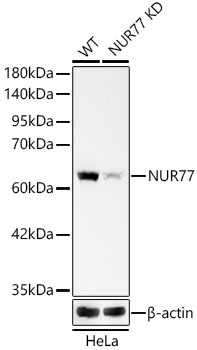
Western blot analysis of lysates from wild type(WT) and NUR77 knockdown (KD) HeLa cells, using [KD Validated] NUR77 Rabbit mAb (CAB24016) at 1:1000 dilution. Secondary antibody: HRP-conjugated Goat anti-Rabbit IgG (H+L) (CABS014) at 1:10000 dilution. Lysates/proteins: 25ug per lane. Blocking buffer: 3% nonfat dry milk in TBST. Detection: ECL Basic Kit (AbGn00020). Exposure time: 10s.
 at 1:1000 dilution. Secondary antibody: HRP Goat Anti-Rabbit IgG (H+L) at 1:10000 dilution. Lysates/proteins: 25ug per lane. Blocking buffer: 3% nonfat dry milk in TBST.")
 at 1:1000 dilution. Secondary antibody: HRP Goat Anti-Rabbit IgG (H+L) at 1:10000 dilution. Lysates/proteins: 25ug per lane. Blocking buffer: 3% nonfat dry milk in TBST.")
