The [KO Validated] KDM1 Antibody (CAB15794) is a high-quality antibody developed for reliable detection and analysis of target proteins. This antibody, produced in rabbits, exhibits high reactivity with human samples and has been validated for use in Western blot applications. By binding specifically to the KDM1A protein, this antibody enables precise detection and analysis of KDM1A levels in various cell types, making it an essential tool for epigenetics and cancer research.KDM1A, also known as lysine-specific demethylase 1A, plays a crucial role in the regulation of gene expression by removing methyl groups from histone proteins.
This antibody is validated for use in WB, IHC-P, IF/ICC, ELISA applications and has demonstrated reactivity against Human, Mouse, Rat samples.
Product Name:
[KO Validated] KDM1 Antibody
SKU:
CAB15794
Size:
20μL, 100μL
Reactivity:
Human, Mouse, Rat
Conjugate:
Unconjugated
Immunogen:
Recombinant protein (or fragment).This information is considered to be commercially sensitive.
Recommended starting concentration is 1 μg/mL. Please optimize the concentration based on your specific assay requirements.
Synonyms:
AOF2, CPRF, KDM1, LSD1, BHC110
Positive Sample:
U-87MG, HeLa, Jurkat, Mouse liver, 293T
Cellular Localization:
Nucleus.
Calculated MW:
93kDa
Observed MW:
110kDa
This gene encodes a nuclear protein containing a SWIRM domain, a FAD-binding motif, and an amine oxidase domain. This protein is a component of several histone deacetylase complexes, though it silences genes by functioning as a histone demethylase. Alternative splicing results in multiple transcript variants.
Purification Method
Affinity purification
Gene ID
23028
RRID
AB_2763215
Buffer Information
Store at -20℃. Avoid freeze / thaw cycles. Buffer: PBS with 0.01% thimerosal,50% glycerol,pH7.3.
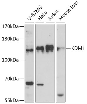
Western blot analysis of various lysates using KDM1 Rabbit pAb (CAB15794) at 1:1000 dilution. Secondary antibody: HRP-conjugated Goat anti-Rabbit IgG (H+L) (CABS014) at 1:10000 dilution. Lysates/proteins: 25μg per lane. Blocking buffer: 3% nonfat dry milk in TBST. Detection: ECL Basic Kit (AbGn00020). Exposure time: 90s.
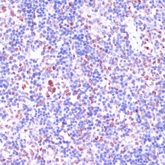
Immunohistochemistry analysis of paraffin-embedded Rat spleen using KDM1 Rabbit pAb (CAB15794) at dilution of 1:100 (40x lens). Microwave antigen retrieval performed with 0.01M PBS Buffer (pH 7.2) prior to IHC staining.
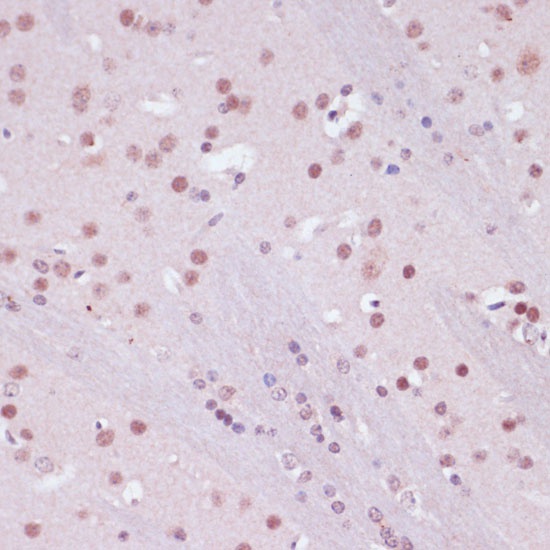
Immunohistochemistry analysis of paraffin-embedded Mouse brain using KDM1 Rabbit pAb (CAB15794) at dilution of 1:100 (40x lens). Microwave antigen retrieval performed with 0.01M PBS Buffer (pH 7.2) prior to IHC staining.
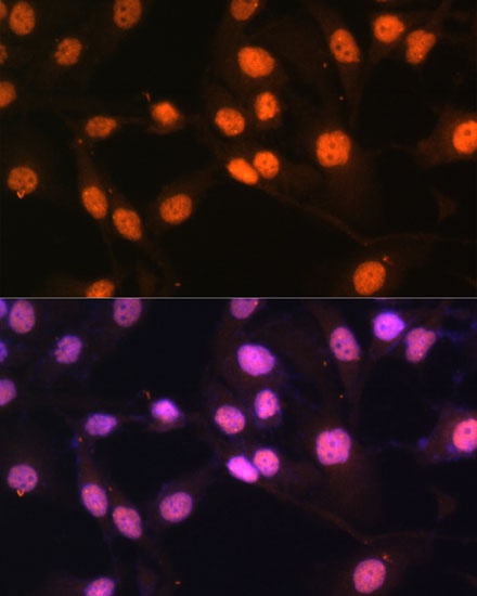
Immunofluorescence analysis of HeLa cells using KDM1 Rabbit pAb (CAB15794) at dilution of 1:100 (40x lens). Secondary antibody: Cy3-conjugated Goat anti-Rabbit IgG (H+L) (CABS007) at 1:500 dilution. Blue: DAPI for nuclear staining.