The KHDRBS1/Sam68 Monoclonal Antibody (CAB3886) is a high-quality antibody developed for reliable detection and analysis of target proteins. This antibody, developed using rabbit monoclonal technology, offers high specificity and sensitivity for detecting SAM68 in human samples. Validated for use in Western blot and immunohistochemistry applications, it allows for precise localization and quantification of SAM68 in different cell types.SAM68 plays a crucial role in regulating alternative splicing, mRNA stability, and translation, making it a key player in gene expression regulation.
This antibody is validated for use in WB, IHC-P, IF/ICC, ELISA applications and has demonstrated reactivity against Human, Mouse, Rat samples.
Product Name:
KHDRBS1/Sam68 Monoclonal Antibody
SKU:
CAB3886
Size:
20μL, 100μL
Reactivity:
Human, Mouse, Rat
Clone Number:
ARC0858
Conjugate:
Unconjugated
Immunogen:
Synthetic peptide. This information is considered to be commercially sensitive.
Recommended starting concentration is 1 μg/mL. Please optimize the concentration based on your specific assay requirements.
Synonyms:
p62, p68, Sam68, KHDRBS1/Sam68
Positive Sample:
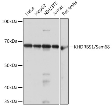
HeLa, HepG2, NIH/3T3, Jurkat, Rat testis
Cellular Localization:
Membrane, Nucleus.
Calculated MW:
48kDa
Observed MW:
68kDa
This gene encodes a member of the K homology domain-containing, RNA-binding, signal transduction-associated protein family. The encoded protein appears to have many functions and may be involved in a variety of cellular processes, including alternative splicing, cell cycle regulation, RNA 3'-end formation, tumorigenesis, and regulation of human immunodeficiency virus gene expression. Alternative splicing results in multiple transcript variants.
Purification Method
Affinity purification
Gene ID
10657
RRID
AB_2863153
Buffer Information
Store at -20℃. Avoid freeze / thaw cycles. Buffer: PBS containing 50% glycerol and 0.05% BSA, preserved with proclin300 or sodium azide, pH 7.3.
Western blot analysis of various lysates using KHDRBS1/KHDRBS1/Sam68 Rabbit mAb (CAB3886) at 1:1000 dilution. Secondary antibody: HRP-conjugated Goat anti-Rabbit IgG (H+L) (CABS014) at 1:10000 dilution. Lysates/proteins: 25μg per lane. Blocking buffer: 3% nonfat dry milk in TBST. Detection: ECL Basic Kit (AbGn00020). Exposure time: 1s.
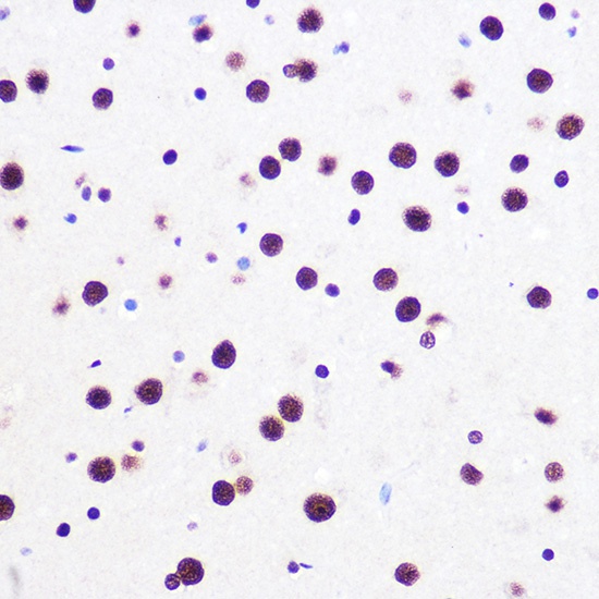
Immunohistochemistry analysis of paraffin-embedded Rat brain using KHDRBS1/KHDRBS1/Sam68 Rabbit mAb (CAB3886) at dilution of 1:100 (40x lens). Microwave antigen retrieval performed with 0.01M PBS Buffer (pH 7.2) prior to IHC staining.
Immunohistochemistry analysis of paraffin-embedded Mouse brain using KHDRBS1/KHDRBS1/Sam68 Rabbit mAb (CAB3886) at dilution of 1:100 (40x lens). Microwave antigen retrieval performed with 0.01M PBS Buffer (pH 7.2) prior to IHC staining.
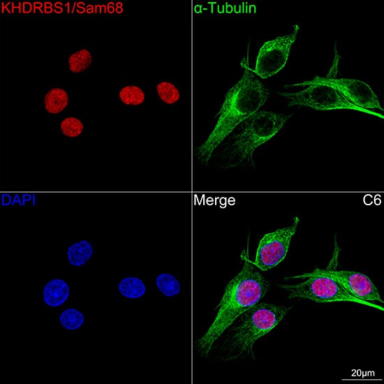
Confocal imaging of C6 cells using KHDRBS1/Sam68 Rabbit mAb (CAB3886,at dilution of 1:100) (Red). The cells were counterstained with α-Tubulin Mouse mAb (AC012,dilution 1:400) (Green). DAPI was used for nuclear staining (blue). Objective: 100x.
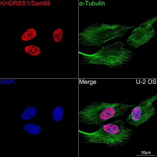
Confocal imaging of U-2 OS cells using KHDRBS1/Sam68 Rabbit mAb (CAB3886,at dilution of 1:100) (Red). The cells were counterstained with α-Tubulin Mouse mAb (AC012,dilution 1:400) (Green). DAPI was used for nuclear staining (blue). Objective: 100x.