The KIFC1 Monoclonal Antibody (CAB0077) is a high-quality antibody developed for reliable detection and analysis of target proteins. This antibody, produced in mice, exhibits high specificity and sensitivity towards KIFC1 in human samples, making it suitable for applications such as immunofluorescence and immunohistochemistry.KIFC1, also known as kinesin-like protein KIFC1, plays a crucial role in mitosis by facilitating the movement of chromosomes during cell division.
This antibody is validated for use in WB, IHC-P, IF/ICC, ELISA applications and has demonstrated reactivity against Human, Mouse samples.
Product Name:
KIFC1 Monoclonal Antibody
SKU:
CAB0077
Size:
20μL, 100μL
Reactivity:
Human, Mouse
Clone Number:
ARC1808
Conjugate:
Unconjugated
Immunogen:
Synthetic peptide. This information is considered to be commercially sensitive.
Recommended starting concentration is 1 μg/mL. Please optimize the concentration based on your specific assay requirements.
Synonyms:
HSET, KNSL2, KIFC1
Positive Sample:
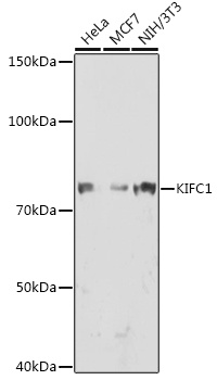
HeLa, MCF7, NIH/3T3
Cellular Localization:
Early Endosome, Microtubule Organizing Center, Mitotic Spindle, Nucleus.
Calculated MW:
74kDa
Observed MW:
74kDa
Predicted to enable microtubule binding activity and minus-end-directed microtubule motor activity. Involved in mitotic metaphase plate congression and mitotic spindle assembly. Located in membrane.
Purification Method
Affinity purification
Gene ID
3833
Buffer Information
Store at -20℃. Avoid freeze / thaw cycles. Buffer: PBS containing 50% glycerol and 0.05% BSA, preserved with proclin300 or sodium azide, pH 7.3.
Western blot analysis of various lysates using KIFC1 Rabbit mAb (CAB0077) at 1:1000 dilution. Secondary antibody: HRP-conjugated Goat anti-Rabbit IgG (H+L) (CABS014) at 1:10000 dilution. Lysates/proteins: 25μg per lane. Blocking buffer: 3% nonfat dry milk in TBST. Detection: ECL Enhanced Kit (AbGn00021). Exposure time: 90s.
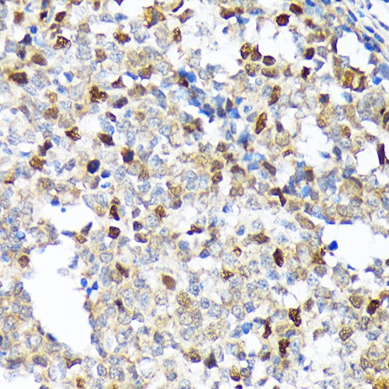
Immunohistochemistry analysis of paraffin-embedded Human appendix using KIFC1 Rabbit mAb (CAB0077) at dilution of 1:100 (40x lens). Microwave antigen retrieval performed with 0.01M Tris/EDTA Buffer (pH 9.0) prior to IHC staining.
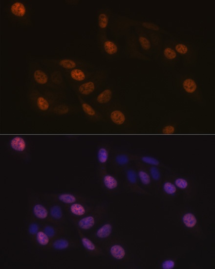
Immunofluorescence analysis of U-2 OS cells using KIFC1 Rabbit mAb (CAB0077) at dilution of 1:100 (40x lens). Secondary antibody: Cy3-conjugated Goat anti-Rabbit IgG (H+L) (CABS007) at 1:500 dilution. Blue: DAPI for nuclear staining.