The KLF4 Antibody (CAB6640) is a high-quality antibody developed for reliable detection and analysis of target proteins. This antibody, produced in rabbits, exhibits high reactivity with human samples and is validated for use in Western blot applications.KLF4, a key player in cellular differentiation and pluripotency, has been linked to various biological processes such as embryonic development, stem cell maintenance, and tumorigenesis. By specifically binding to the KLF4 protein, this antibody enables accurate detection and analysis in different cell types, offering valuable insights for studies in stem cell biology, cancer research, and developmental biology.
This antibody is validated for use in WB, IHC-P, IF/ICC, ELISA applications and has demonstrated reactivity against Human, Mouse, Rat samples.
Product Name:
KLF4 Antibody
SKU:
CAB6640
Size:
20μL, 100μL
Reactivity:
Human, Mouse, Rat
Conjugate:
Unconjugated
Immunogen:
Recombinant protein (or fragment).This information is considered to be commercially sensitive.
Recommended starting concentration is 1 μg/mL. Please optimize the concentration based on your specific assay requirements.
Synonyms:
EZF, GKLF, KLF4
Positive Sample:
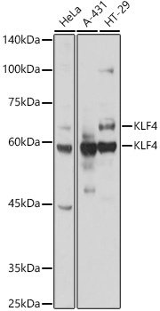
HeLa, HCT116, HT-29
Cellular Localization:
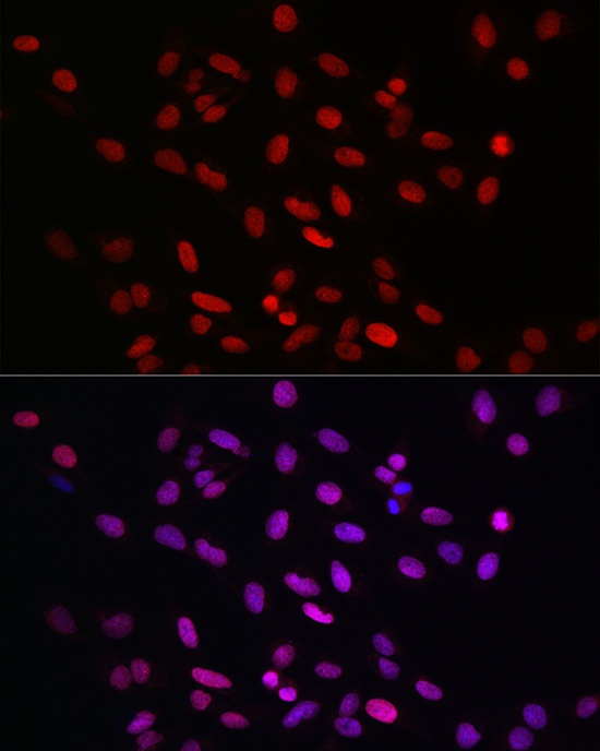
Nucleus.
Calculated MW:
55kDa
Observed MW:
62kDa
This gene encodes a protein that belongs to the Kruppel family of transcription factors. The encoded zinc finger protein is required for normal development of the barrier function of skin. The encoded protein is thought to control the G1-to-S transition of the cell cycle following DNA damage by mediating the tumor suppressor gene p53. Mice lacking this gene have a normal appearance but lose weight rapidly, and die shortly after birth due to fluid evaporation resulting from compromised epidermal barrier function. Alternative splicing results in multiple transcript variants encoding different isoforms.
Purification Method
Affinity purification
Gene ID
9314
RRID
AB_2767229
Buffer Information
Store at -20℃. Avoid freeze / thaw cycles. Buffer: PBS containing 50% glycerol, preserved with proclin300 or sodium azide, pH 7.3.
Western blot analysis of various lysates, using KLF4 Rabbit pAb (CAB6640) at 1:1000 dilution. Secondary antibody: HRP-conjugated Goat anti-Rabbit IgG (H+L) (CABS014) at 1:10000 dilution. Lysates/proteins: 25μg per lane. Blocking buffer: 3% nonfat dry milk in TBST. Detection: ECL Basic Kit (AbGn00020). Exposure time: 30s.
Western blot analysis of various lysates, using KLF4 Rabbit pAb (CAB6640) at 1:1000 dilution. Secondary antibody: HRP-conjugated Goat anti-Rabbit IgG (H+L) (CABS014) at 1:10000 dilution. Lysates/proteins: 25μg per lane. Blocking buffer: 3% nonfat dry milk in TBST. Detection: ECL Basic Kit (AbGn00020). Exposure time: 90s.
Immunofluorescence analysis of U2OS cells using KLF4 Rabbit pAb (CAB6640) at dilution of 1:100 (40x lens). Secondary antibody: Cy3-conjugated Goat anti-Rabbit IgG (H+L) (CABS007) at 1:500 dilution. Blue: DAPI for nuclear staining.