The [KO Validated] AK2 Antibody (CAB6519) is a high-quality antibody developed for reliable detection and analysis of target proteins. This antibody, raised in rabbits, has been validated for use in various research applications, including Western blot and immunohistochemistry.AK2 is a crucial protein that plays a vital role in the maintenance of mitochondrial function and energy production within cells. Dysregulation of AK2 has been linked to various diseases, including metabolic disorders, neurodegenerative diseases, and cancer. Therefore, studying AK2 expression and activity is essential for understanding its impact on cellular physiology and disease progression.
This antibody is validated for use in WB, IHC-P, ELISA applications and has demonstrated reactivity against Human, Mouse, Rat samples.
Product Name:
[KO Validated] AK2 Antibody
SKU:
CAB6519
Size:
20μL, 100μL
Reactivity:
Human, Mouse, Rat
Conjugate:
Unconjugated
Immunogen:
Recombinant protein (or fragment).This information is considered to be commercially sensitive.
Recommended starting concentration is 1 μg/mL. Please optimize the concentration based on your specific assay requirements.
Synonyms:
ADK2, K2
Positive Sample:
HL-60, Mouse liver, Mouse kidney, Rat liver
Cellular Localization:
Mitochondrion Intermembrane Space.
Calculated MW:
26kDa
Observed MW:
32kDa
Adenylate kinases are involved in regulating the adenine nucleotide composition within a cell by catalyzing the reversible transfer of phosphate groups among adenine nucleotides. Three isozymes of adenylate kinase, namely 1, 2, and 3, have been identified in vertebrates; this gene encodes isozyme 2. Expression of these isozymes is tissue-specific and developmentally regulated. Isozyme 2 is localized in the mitochondrial intermembrane space and may play a role in apoptosis. Mutations in this gene are the cause of reticular dysgenesis. Alternate splicing results in multiple transcript variants. Pseudogenes of this gene are found on chromosomes 1 and 2.
Purification Method
Affinity purification
Gene ID
204
RRID
AB_2863530
Buffer Information
Store at -20℃. Avoid freeze / thaw cycles. Buffer: PBS containing 50% glycerol, preserved with proclin300 or sodium azide, pH 7.3.
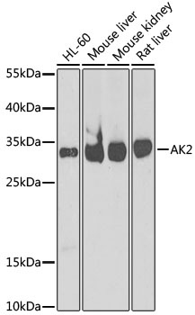
Western blot analysis of various lysates using [KO Validated] AK2 Rabbit pAb (CAB6519) at 1:1000 dilution. Secondary antibody: HRP-conjugated Goat anti-Rabbit IgG (H+L) (CABS014) at 1:10000 dilution. Lysates/proteins: 25μg per lane. Blocking buffer: 3% nonfat dry milk in TBST. Detection: ECL Basic Kit (AbGn00020). Exposure time: 90s.
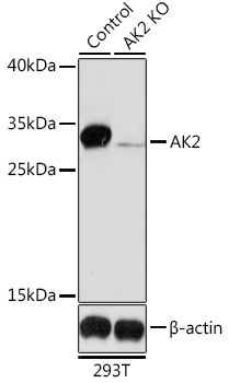
Western blot analysis of lysates from wild type (WT) and AK2 knockout (KO) 293T cells, using [KO Validated] AK2 Rabbit pAb (CAB6519) at 1:1000 dilution. Secondary antibody: HRP-conjugated Goat anti-Rabbit IgG (H+L) (CABS014) at 1:10000 dilution. Lysates/proteins: 25μg per lane. Blocking buffer: 3% nonfat dry milk in TBST. Detection: ECL Basic Kit (AbGn00020). Exposure time: 90s.