The [KO Validated] ANXA1 Antibody (CAB1118) is a high-quality antibody developed for reliable detection and analysis of target proteins. This antibody, produced in rabbits, is highly specific for human Annexin A1 and has been validated for use in Western blot applications.Annexin A1 is a key player in the resolution of inflammation, acting as an anti-inflammatory mediator by inhibiting leukocyte migration and promoting the clearance of apoptotic cells. Dysregulation of Annexin A1 has been implicated in various inflammatory disorders, making it a subject of interest in research on conditions such as arthritis, asthma, and cancer.
This antibody is validated for use in WB, IHC-P, IF/ICC, ELISA applications and has demonstrated reactivity against Human, Mouse, Rat samples.
Product Name:
[KO Validated] ANXA1 Antibody
SKU:
CAB1118
Size:
20μL, 100μL
Reactivity:
Human, Mouse, Rat
Conjugate:
Unconjugated
Immunogen:
Recombinant protein (or fragment).This information is considered to be commercially sensitive.
This gene encodes a membrane-localized protein that binds phospholipids. This protein inhibits phospholipase A2 and has anti-inflammatory activity. Loss of function or expression of this gene has been detected in multiple tumors.
Purification Method
Affinity purification
Gene ID
301
RRID
AB_2861514
Buffer Information
Store at -20℃. Avoid freeze / thaw cycles. Buffer: PBS containing 50% glycerol, preserved with proclin300 or sodium azide, pH 7.3.
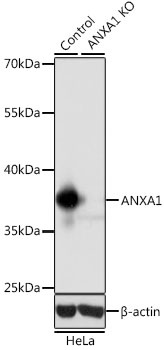
Western blot analysis of lysates from wild type (WT) and ANXA1 knockout (KO) HeLa cells, using [KO Validated] ANXA1 Rabbit pAb (CAB1118) at 1:1000 dilution. Secondary antibody: HRP-conjugated Goat anti-Rabbit IgG (H+L) (CABS014) at 1:10000 dilution. Lysates/proteins: 25μg per lane. Blocking buffer: 3% nonfat dry milk in TBST. Detection: ECL Basic Kit (AbGn00020). Exposure time: 1s.
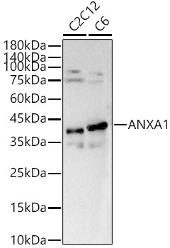
Western blot analysis of various lysates, using [KO Validated] ANXA1 Rabbit pAb (CAB1118) at 1:700 dilution. Secondary antibody: HRP-conjugated Goat anti-Rabbit IgG (H+L) (CABS014) at 1:10000 dilution. Lysates/proteins: 25μg per lane. Blocking buffer: 3% nonfat dry milk in TBST. Detection: ECL Basic Kit (AbGn00020). Exposure time: 90s.
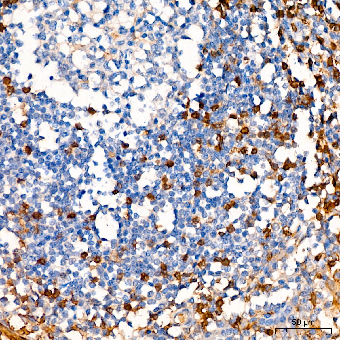
Immunohistochemistry analysis of paraffin-embedded Human tonsil using [KO Validated] ANXA1 Rabbit pAb (CAB1118) at dilution of 1:50 (40x lens). High pressure antigen retrieval performed with 0.01M Citrate buffer (pH 6.0) prior to IHC staining.
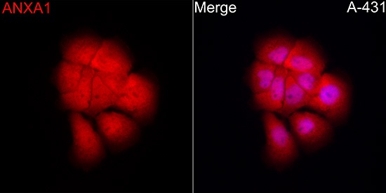
Immunofluorescence analysis of A-431 cells using [KO Validated] ANXA1 Rabbit pAb (CAB1118) at dilution of 1:100 (40x lens). Secondary antibody: Cy3-conjugated Goat anti-Rabbit IgG (H+L) (CABS007) at 1:500 dilution. Blue: DAPI for nuclear staining.
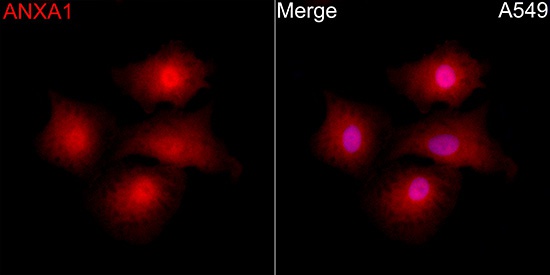
Immunofluorescence analysis of A-549 cells using [KO Validated] ANXA1 Rabbit pAb (CAB1118) at dilution of 1:100 (40x lens). Secondary antibody: Cy3-conjugated Goat anti-Rabbit IgG (H+L) (CABS007) at 1:500 dilution. Blue: DAPI for nuclear staining.
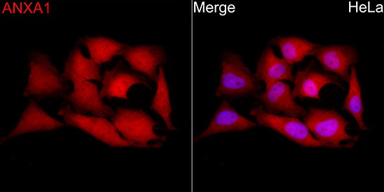
Immunofluorescence analysis of HeLa cells using [KO Validated] ANXA1 Rabbit pAb (CAB1118) at dilution of 1:100 (40x lens). Secondary antibody: Cy3-conjugated Goat anti-Rabbit IgG (H+L) (CABS007) at 1:500 dilution. Blue: DAPI for nuclear staining.