The [KO Validated] APEX1/APE1 Antibody (CAB2587) is a high-quality antibody developed for reliable detection and analysis of target proteins. This antibody, produced in rabbits, exhibits high reactivity with human samples and has been rigorously validated for Western blot applications.APEX1, also known as redox factor-1, plays a crucial role in maintaining genomic integrity and cellular homeostasis by repairing DNA damage caused by oxidative stress. Its involvement in these essential processes makes it a key target for research in fields such as cancer biology, neurodegeneration, and aging.
This antibody is validated for use in WB, IF/ICC, IP, ELISA applications and has demonstrated reactivity against Human samples.
Product Name:
[KO Validated] APEX1/APE1 Antibody
SKU:
CAB2587
Size:
20μL, 100μL
Reactivity:
Human
Conjugate:
Unconjugated
Immunogen:
Recombinant protein (or fragment).This information is considered to be commercially sensitive.
The APEX gene encodes the major AP endonuclease in human cells. It encodes the APEX endonuclease, a DNA repair enzyme with apurinic/apyrimidinic (AP) activity. Such AP activity sites occur frequently in DNA molecules by spontaneous hydrolysis, by DNA damaging agents or by DNA glycosylases that remove specific abnormal bases. The AP sites are the most frequent pre-mutagenic lesions that can prevent normal DNA replication. Splice variants have been found for this gene; all encode the same protein. Disruptions in the biological functions related to APEX are associated with many various malignancies and neurodegenerative diseases.
Purification Method
Affinity purification
Gene ID
328
RRID
AB_2863015
Buffer Information
Store at -20℃. Avoid freeze / thaw cycles. Buffer: PBS containing 50% glycerol, preserved with proclin300 or sodium azide, pH 7.3.
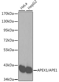
Western blot analysis of various lysates using [KO Validated] APEX1/APE1 Rabbit pAb (CAB2587). Secondary antibody: HRP-conjugated Goat anti-Rabbit IgG (H+L) (CABS014) at 1:10000 dilution. Lysates/proteins: 25μg per lane. Blocking buffer: 3% nonfat dry milk in TBST.
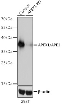
Western blot analysis of lysates from wild type (WT) and APEX1/APE1 knockout (KO) 293T cells, using [KO Validated] APEX1/APE1 Rabbit pAb (CAB2587) at 1:3000 dilution. Secondary antibody: HRP-conjugated Goat anti-Rabbit IgG (H+L) (CABS014) at 1:10000 dilution. Lysates/proteins: 25μg per lane. Blocking buffer: 3% nonfat dry milk in TBST. Detection: ECL Basic Kit (AbGn00020). Exposure time: 1s.
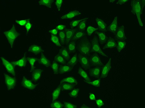
Immunofluorescence analysis of A549 cells using [KO Validated] APEX1/APE1 Rabbit pAb (CAB2587).Secondary antibody: Cy3-conjugated Goat anti-Rabbit IgG (H+L) (CABS007) at 1:500 dilution.
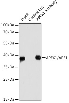
Immunoprecipitation analysis of 200 μg extracts of HeLa cells using 1 μg APEX1/APE1 antibody (CAB2587). Western blot was performed from the immunoprecipitate using APEX1/APE1 antibody (CAB2587) at a dilution of 1:1000.