The [KO Validated] CDK4 Monoclonal Antibody (CAB11136) is a high-quality antibody developed for reliable detection and analysis of target proteins. The protein encoded by this gene is a member of the Ser/Thr protein kinase family. This protein is highly similar to the gene products of S. cerevisiae cdc28 and S. pombe cdc2. It is a catalytic subunit of the protein kinase complex that is important for cell cycle G1 phase progression. The activity of this kinase is restricted to the G1-S phase, which is controlled by the regulatory subunits D-type cyclins and CDK inhibitor p16(INK4a). This kinase was shown to be responsible for the phosphorylation of retinoblastoma gene product (Rb). Mutations in this gene as well as in its related proteins including D-type cyclins, p16(INK4a) and Rb were all found to be associated with tumorigenesis of a variety of cancers. Multiple polyadenylation sites of this gene have been reported.
This antibody is validated for use in WB, IHC-P, IP, ELISA applications and has demonstrated reactivity against Human, Mouse, Rat, Monkey samples.
Product Name:
[KO Validated] CDK4 Monoclonal Antibody
SKU:
CAB11136
Size:
100μL, 20μL
Reactivity:
Human, Mouse, Rat, Monkey
Clone Number:
ARC51004
Conjugate:
Unconjugated
Immunogen:
Synthetic peptide. This information is considered to be commercially sensitive.
Tested Applications:
WBIHC-PIPELISA
Recommended Dilution:
WB
1:1000 - 1:6000
IP
0.5μg-4μg antibody for 200μg-400μg extracts of whole cells
IHC-P
1:200 - 1:2000
ELISA
Recommended starting concentration is 1 μg/mL. Please optimize the concentration based on your specific assay requirements.For high-ratio antibody dilutions (≥1:10000),a sequential dilution method is strongly recommended to ensure measurement accuracy.
Synonyms:
CMM3, PSK-J3, K4
Positive Sample:
HeLa, COS-7, Mouse lung, Rat lung, NIH/3T3
Cellular Localization:
Cytoplasm, Membrane, Nucleus.
Calculated MW:
34kDa
Observed MW:
34kDa
The protein encoded by this gene is a member of the Ser/Thr protein kinase family. This protein is highly similar to the gene products of S. cerevisiae cdc28 and S. pombe cdc2. It is a catalytic subunit of the protein kinase complex that is important for cell cycle G1 phase progression. The activity of this kinase is restricted to the G1-S phase, which is controlled by the regulatory subunits D-type cyclins and CDK inhibitor p16(INK4a). This kinase was shown to be responsible for the phosphorylation of retinoblastoma gene product (Rb). Mutations in this gene as well as in its related proteins including D-type cyclins, p16(INK4a) and Rb were all found to be associated with tumorigenesis of a variety of cancers. Multiple polyadenylation sites of this gene have been reported.
Purification Method
Affinity purification
Gene ID
1019
RRID
AB_2861505
Buffer Information
Store at -20℃. Avoid freeze / thaw cycles. Buffer: PBS containing 50% glycerol and 0.05% BSA, preserved with proclin300 or sodium azide, pH 7.3.
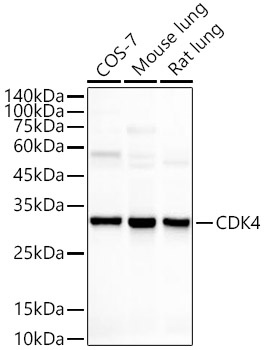
Western blot analysis of various lysates, using [KO Validated] CDK4 Rabbit mAb (CAB11136) at 1:1000 dilution. Secondary antibody: HRP-conjugated Goat anti-Rabbit IgG (H+L) (AS014) at 1:10000 dilution. Lysates/proteins: 25μg per lane. Blocking buffer: 3% nonfat dry milk in TBST. Detection: ECL Basic Kit (AbGn00020). Exposure time: 10s.
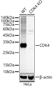
Western blot analysis of lysates from wild type(WT) and knockout (KO) HeLa cells, using [KO Validated] CDK4 Rabbit mAb (CAB11136) at 1:1000 dilution. Secondary antibody: HRP-conjugated Goat anti-Rabbit IgG (H+L) (AS014) at 1:10000 dilution. Lysates/proteins: 25μg per lane. Blocking buffer: 3% nonfat dry milk in TBST. Detection: ECL Basic Kit (AbGn00020). Exposure time: 10s.
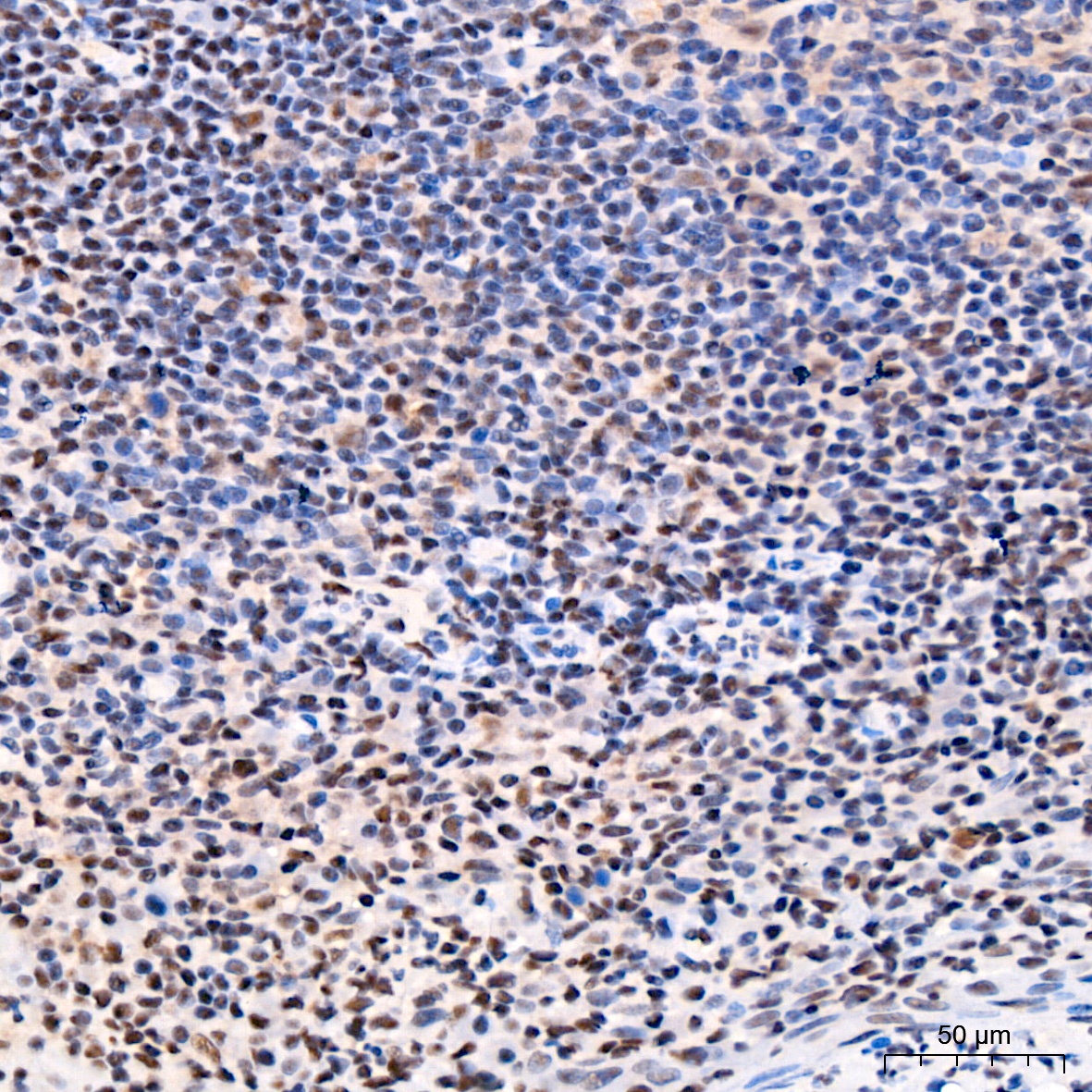
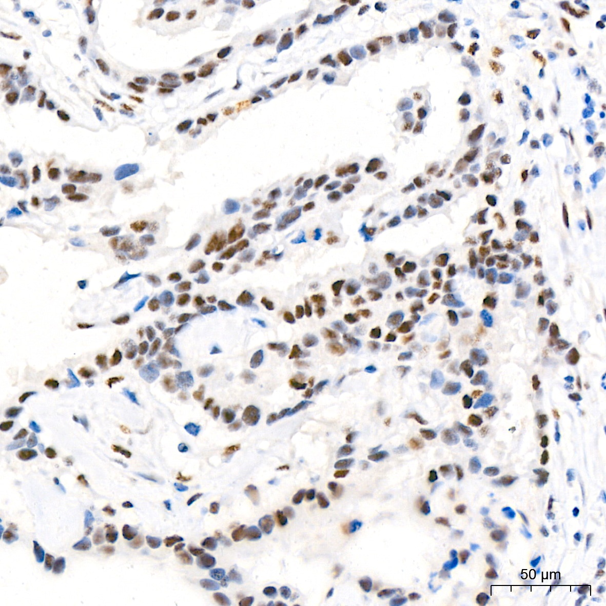
Immunohistochemistry analysis of paraffin-embedded Human tonsil tissue using [KO Validated] CDK4 Rabbit mAb (CAB11136) at a dilution of 1:500 (40x lens). High pressure antigen retrieval performed with 0.01M Citrate buffer (pH 6.0) prior to IHC staining.
Immunohistochemistry analysis of paraffin-embedded Human breast cancer tissue using [KO Validated] CDK4 Rabbit mAb (CAB11136) at a dilution of 1:500 (40x lens). High pressure antigen retrieval performed with 0.01M Citrate buffer (pH 6.0) prior to IHC staining.
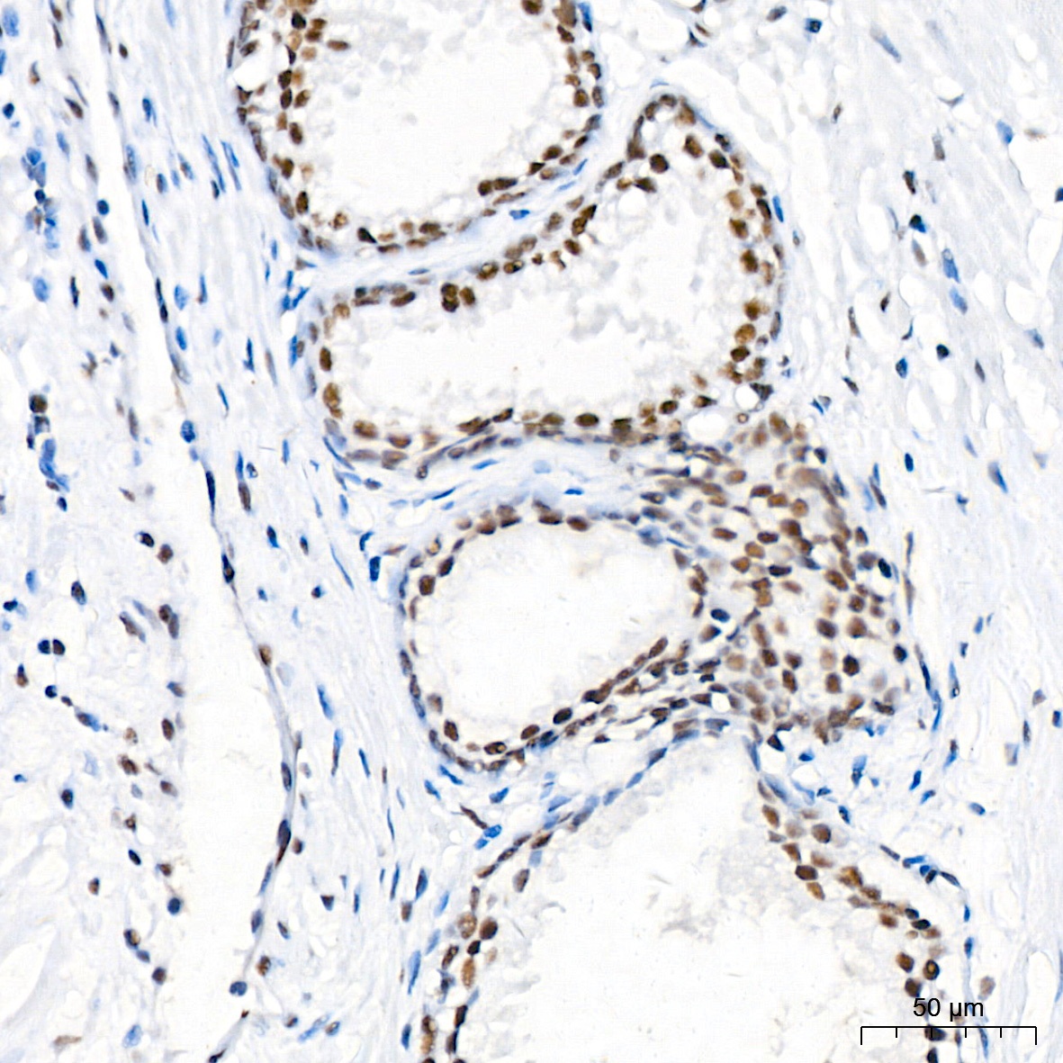
Immunohistochemistry analysis of paraffin-embedded Human prostate tissue using [KO Validated] CDK4 Rabbit mAb (CAB11136) at a dilution of 1:500 (40x lens). High pressure antigen retrieval performed with 0.01M Citrate buffer (pH 6.0) prior to IHC staining.
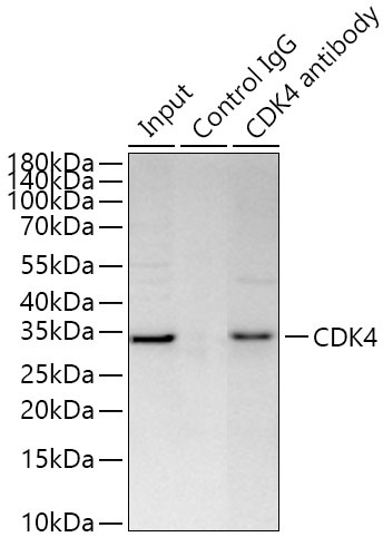
Immunoprecipitation of CDK4 from 300 µg extracts of HeLa was performed using 0.5 µg of [KO Validated] CDK4 Rabbit mAb (CAB11136). Rabbit IgG isotype control (AC005) was used to precipitate the Control IgG sample. IP samples were eluted with 1X reducing Laemmli Buffer. The Input lane represents 10% of the total input. Western blot analysis of immunoprecipitates was conducted using [KO Validated] CDK4 Rabbit mAb (CAB11136) at a dilution of 1:1000.