The [KO Validated] EGFR Antibody (CAB11351) is a high-quality antibody developed for reliable detection and analysis of target proteins. This antibody, produced in rabbits, has been extensively validated for its specificity and sensitivity in various research applications, including Western blotting.EGFR is a transmembrane tyrosine kinase receptor that plays a crucial role in regulating cell growth and differentiation. Dysregulation of EGFR signaling is commonly observed in various cancers, making it an attractive target for cancer therapy. The KO-Validated EGFR Polyclonal Antibody specifically binds to EGFR, enabling researchers to detect and analyze its expression in different cell types and tissues.
This antibody is validated for use in WB, IHC-P, IF/ICC, IP, ELISA applications and has demonstrated reactivity against Human, Mouse, Rat samples.
Product Name:
[KO Validated] EGFR Antibody
SKU:
CAB11351
Size:
20μL, 100μL
Reactivity:
Human, Mouse, Rat
Conjugate:
Unconjugated
Immunogen:
Recombinant protein (or fragment).This information is considered to be commercially sensitive.
Cell Membrane, Endoplasmic Reticulum Membrane, Endosome, Endosome Membrane, Golgi Apparatus Membrane, Nucleus Membrane, Nucleus, Secreted, Single-Pass Type I Membrane Protein.
Calculated MW:
134kDa
Observed MW:
175kDa
The protein encoded by this gene is a transmembrane glycoprotein that is a member of the protein kinase superfamily. This protein is a receptor for members of the epidermal growth factor family. EGFR is a cell surface protein that binds to epidermal growth factor, thus inducing receptor dimerization and tyrosine autophosphorylation leading to cell proliferation. Mutations in this gene are associated with lung cancer. EGFR is a component of the cytokine storm which contributes to a severe form of Coronavirus Disease 2019 (COVID-19) resulting from infection with severe acute respiratory syndrome coronavirus-2 (SARS-CoV-2).
Purification Method
Affinity purification
Gene ID
1956
RRID
AB_2861549
Buffer Information
Store at -20℃. Avoid freeze / thaw cycles. Buffer: PBS containing 50% glycerol, preserved with proclin300 or sodium azide, pH 7.3.
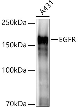
Western blot analysis of lysates from A431 cells, using EGFR Rabbit pAb (CAB11351) at 1:2000 dilution. Secondary antibody: HRP-conjugated Goat anti-Rabbit IgG (H+L) (CABS014) at 1:10000 dilution. Lysates/proteins: 25μg per lane. Blocking buffer: 3% nonfat dry milk in TBST. Detection: ECL Basic Kit (AbGn00020). Exposure time: 3s.
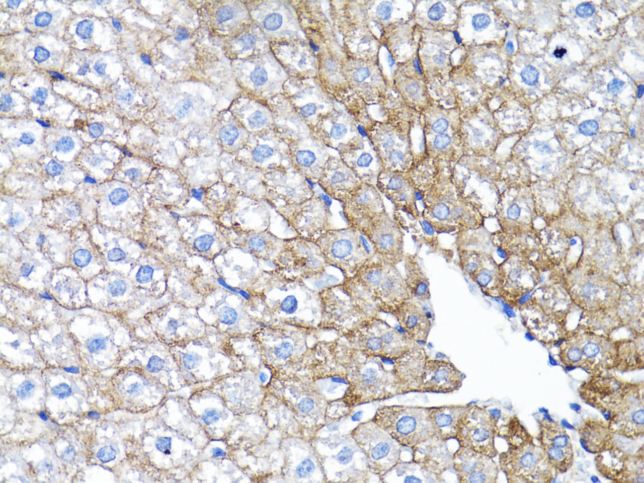
Immunohistochemistry analysis of paraffin-embedded Rat liver using EGFR Rabbit pAb (CAB11351) at dilution of 1:200 (40x lens). Microwave antigen retrieval performed with 0.01M PBS Buffer (pH 7.2) prior to IHC staining.
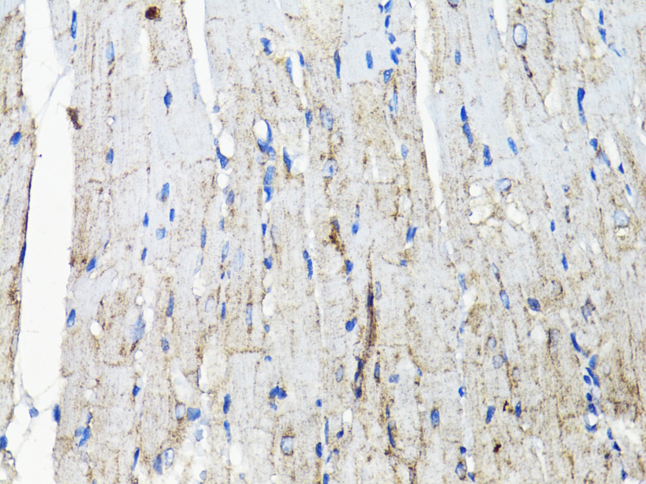
Immunohistochemistry analysis of paraffin-embedded Mouse heart using EGFR Rabbit pAb (CAB11351) at dilution of 1:200 (40x lens). Microwave antigen retrieval performed with 0.01M PBS Buffer (pH 7.2) prior to IHC staining.
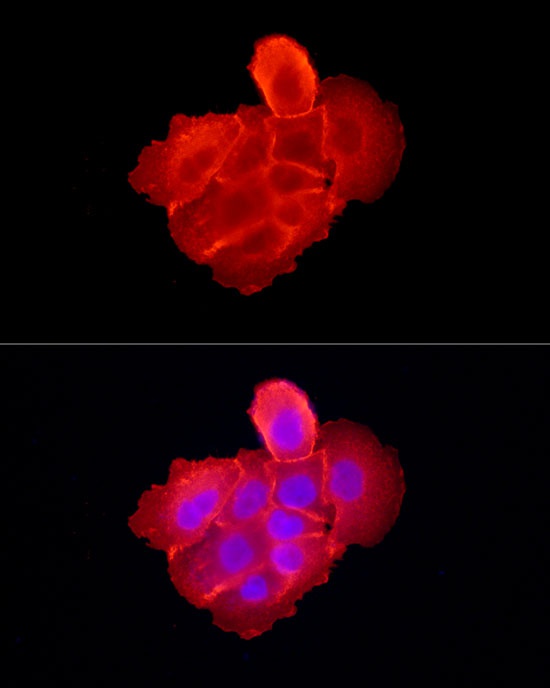
Immunofluorescence analysis of A-431 cells using EGFR Rabbit pAb (CAB11351) at dilution of 1:300 (40x lens). Secondary antibody: Cy3-conjugated Goat anti-Rabbit IgG (H+L) (CABS007) at 1:500 dilution. Blue: DAPI for nuclear staining.