The [KO Validated] Fascin/FSCN1 Antibody (CAB1904) is a high-quality antibody developed for reliable detection and analysis of target proteins. This gene encodes a member of the fascin family of actin-binding proteins. Fascin proteins organize F-actin into parallel bundles, and are required for the formation of actin-based cellular protrusions. The encoded protein plays a critical role in cell migration, motility, adhesion and cellular interactions. Expression of this gene is known to be regulated by several microRNAs, and overexpression of this gene may play a role in the metastasis of multiple types of cancer by increasing cell motility. Expression of this gene is also a marker for Reed-Sternberg cells in Hodgkin's lymphoma. A pseudogene of this gene is located on the long arm of chromosome 15.
This antibody is validated for use in WB, IHC-P, IF/ICC, ELISA applications and has demonstrated reactivity against Human, Mouse, Rat samples.
Product Name:
[KO Validated] Fascin/FSCN1 Antibody
SKU:
CAB1904
Size:
100μL, 20μL
Reactivity:
Human, Mouse, Rat
Conjugate:
Unconjugated
Immunogen:
Recombinant protein (or fragment).This information is considered to be commercially sensitive.
Tested Applications:
WBIHC-PIF/ICCELISA
Recommended Dilution:
WB
1:500 - 1:2000
IHC-P
1:50 - 1:200
IF/ICC
1:50 - 1:200
ELISA
Recommended starting concentration is 1 μg/mL. Please optimize the concentration based on your specific assay requirements.
This gene encodes a member of the fascin family of actin-binding proteins. Fascin proteins organize F-actin into parallel bundles, and are required for the formation of actin-based cellular protrusions. The encoded protein plays a critical role in cell migration, motility, adhesion and cellular interactions. Expression of this gene is known to be regulated by several microRNAs, and overexpression of this gene may play a role in the metastasis of multiple types of cancer by increasing cell motility. Expression of this gene is also a marker for Reed-Sternberg cells in Hodgkin's lymphoma. A pseudogene of this gene is located on the long arm of chromosome 15.
Purification Method
Affinity purification
Gene ID
6624
RRID
AB_2862532
Buffer Information
Store at -20℃. Avoid freeze / thaw cycles. Buffer: PBS containing 50% glycerol, preserved with proclin300 or sodium azide, pH 7.3.
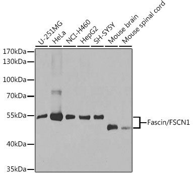
Western blot analysis of various lysates using [KO Validated] Fascin/FSCN1 Rabbit pAb (CAB1904) at 1:1000 dilution. Secondary antibody: HRP-conjugated Goat anti-Rabbit IgG (H+L) (AS014) at 1:10000 dilution. Lysates/proteins: 25μg per lane. Blocking buffer: 3% nonfat dry milk in TBST. Detection: ECL Basic Kit (AbGn00020). Exposure time: 30s.
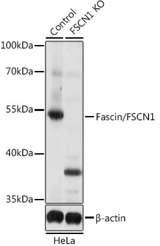
Western blot analysis of lysates from wild type (WT) and Fascin/Fascin/FSCN1 knockout (KO) HeLa cells, using [KO Validated] Fascin/FSCN1 Rabbit pAb (CAB1904) at 1:1000 dilution. Secondary antibody: HRP-conjugated Goat anti-Rabbit IgG (H+L) (AS014) at 1:10000 dilution. Lysates/proteins: 25μg per lane. Blocking buffer: 3% nonfat dry milk in TBST. Detection: ECL Basic Kit (AbGn00020). Exposure time: 5s.
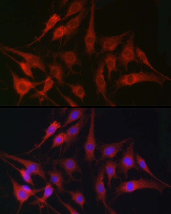
Immunofluorescence analysis of C6 cells using [KO Validated] Fascin/Fascin/FSCN1 Rabbit pAb (CAB1904) at dilution of 1:100 (40x lens). Secondary antibody: Cy3-conjugated Goat anti-Rabbit IgG (H+L) (AS007) at 1:500 dilution. Blue: DAPI for nuclear staining.
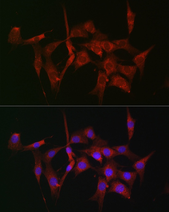
Immunofluorescence analysis of NIH-3T3 cells using [KO Validated] Fascin/Fascin/FSCN1 Rabbit pAb (CAB1904) at dilution of 1:100 (40x lens). Secondary antibody: Cy3-conjugated Goat anti-Rabbit IgG (H+L) (AS007) at 1:500 dilution. Blue: DAPI for nuclear staining.