The [KO Validated] Lamin B1 Antibody (CAB1910) is a high-quality antibody developed for reliable detection and analysis of target proteins. This antibody, produced through immunization of rabbits, is highly specific and reactive with human samples, making it ideal for use in Western blot and immunohistochemistry applications.Lamin B1 is a crucial component of the nuclear lamina, providing structural support to the nucleus and playing a role in various cellular processes such as DNA replication, transcription, and nuclear envelope stability. Dysregulation of Lamin B1 has been implicated in various diseases, including progeria and certain cancers, making it an important target for biological research.
This antibody is validated for use in WB, IHC-P, IF/ICC, IP, ChIP, ELISA applications and has demonstrated reactivity against Human, Mouse, Rat samples.
Product Name:
[KO Validated] Lamin B1 Antibody
SKU:
CAB1910
Size:
20μL, 100μL
Reactivity:
Human, Mouse, Rat
Conjugate:
Unconjugated
Immunogen:
Recombinant protein (or fragment).This information is considered to be commercially sensitive.
This gene encodes one of the two B-type lamin proteins and is a component of the nuclear lamina. A duplication of this gene is associated with autosomal dominant adult-onset leukodystrophy (ADLD). Alternative splicing results in multiple transcript variants.
Purification Method
Affinity purification
Gene ID
4001
RRID
AB_2862592
Buffer Information
Store at -20℃. Avoid freeze / thaw cycles. Buffer: PBS containing 50% glycerol, preserved with proclin300 or sodium azide, pH 7.3.
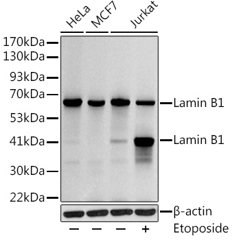
Western blot analysis of various lysates using Lamin B1 Rabbit pAb (CAB1910) at 1:1000 dilution. Jurkat cells were treated with Etoposide (25 uM) at 37℃ for 5 hours. Secondary antibody: HRP-conjugated Goat anti-Rabbit IgG (H+L) (CABS014) at 1:10000 dilution. Lysates/proteins: 25μg per lane. Blocking buffer: 3% nonfat dry milk in TBST. Detection: ECL Basic Kit (AbGn00020). Exposure time: 10s.
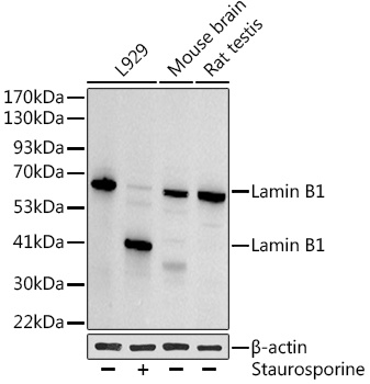
Western blot analysis of various lysates using Lamin B1 Rabbit pAb (CAB1910) at 1:1000 dilution. L929 cells were treated with staurosporine(1 uM) for 3 hour. Secondary antibody: HRP-conjugated Goat anti-Rabbit IgG (H+L) (CABS014) at 1:10000 dilution. Lysates/proteins: 25μg per lane. Blocking buffer: 3% nonfat dry milk in TBST. Detection: ECL Basic Kit (AbGn00020). Exposure time: 30s.
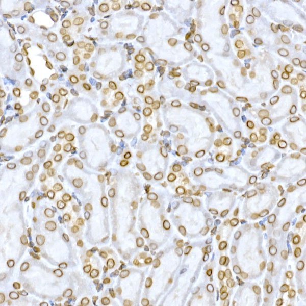
Immunohistochemistry analysis of paraffin-embedded Mouse kidney using Lamin B1 Rabbit pAb (CAB1910) at dilution of 1:150 (40x lens). High pressure antigen retrieval performed with 0.01M Citrate buffer (pH 6.0) prior to IHC staining.
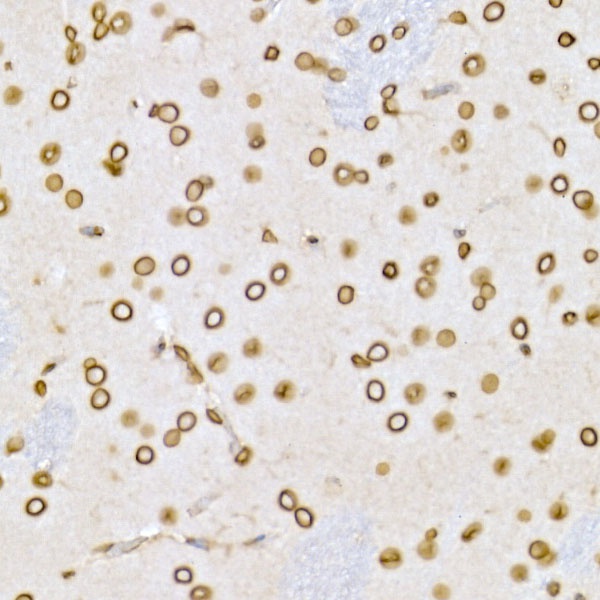
Immunohistochemistry analysis of paraffin-embedded Rat brain using Lamin B1 Rabbit pAb (CAB1910) at dilution of 1:150 (40x lens). High pressure antigen retrieval performed with 0.01M Citrate buffer (pH 6.0) prior to IHC staining.
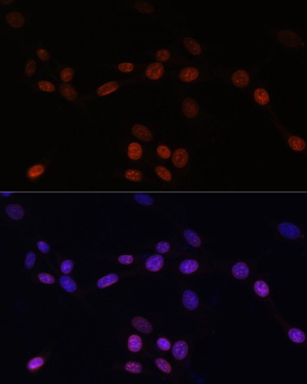
Immunofluorescence analysis of NIH/3T3 cells using Lamin B1 Rabbit pAb (CAB1910) at dilution of 1:100. Secondary antibody: Cy3-conjugated Goat anti-Rabbit IgG (H+L) (CABS007) at 1:500 dilution. Blue: DAPI for nuclear staining.
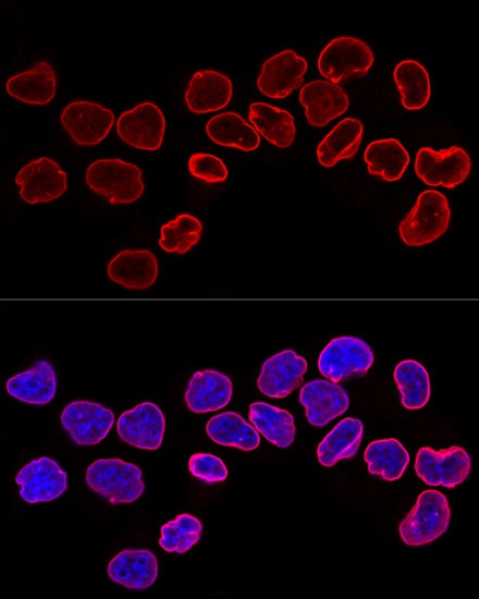
Confocal immunofluorescence analysis of HeLa cells using Lamin B1 Rabbit pAb (CAB1910) at dilution of 1:200. Blue: DAPI for nuclear staining.
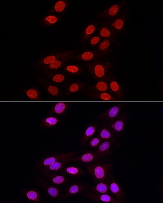
Immunofluorescence analysis of U2OS cells using Lamin B1 Rabbit pAb (CAB1910) at dilution of 1:100 (40x lens). Secondary antibody: Cy3-conjugated Goat anti-Rabbit IgG (H+L) (CABS007) at 1:500 dilution. Blue: DAPI for nuclear staining.
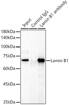
Immunoprecipitation analysis of 300 μg extracts of HeLa cells using 3 μg Lamin B1 antibody (CAB1910). Western blot was performed from the immunoprecipitate using Lamin B1 antibody (CAB1910) at a dilution of 1:1000.
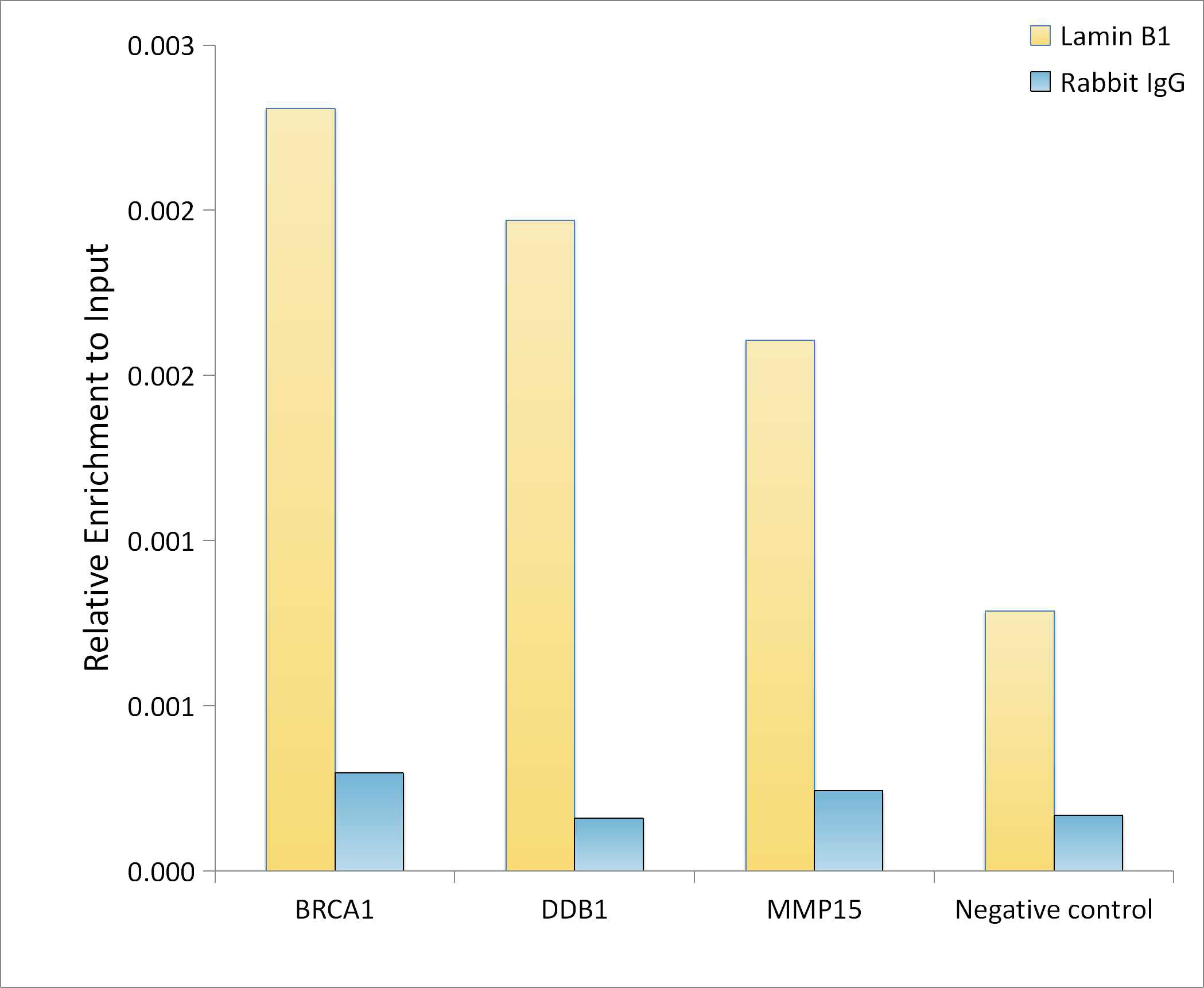
Chromatin immunoprecipitation analysis of extracts of HeLa cells, using Lamin B1 Rabbit pAb antibody (CAB1910) and rabbit IgG.The amount of immunoprecipitated DNA was checked by quantitative PCR. Histogram was constructed by the ratios of the immunoprecipitated DNA to the input.