The [KO Validated] LC3B Monoclonal Antibody (CAB19665) is a high-quality antibody developed for reliable detection and analysis of target proteins. This antibody, generated in rabbits, is highly specific for the LC3B protein, a key marker of autophagosomes, and has been rigorously validated for knockout (KO) studies.LC3B is a critical protein involved in the formation and maturation of autophagosomes, the structures responsible for engulfing and degrading cellular components. Dysregulation of autophagy has been implicated in various diseases, including neurodegenerative disorders, cancer, and metabolic syndromes.
This antibody is validated for use in WB, IHC-P, IF/ICC, IP, ELISA applications and has demonstrated reactivity against Human, Mouse, Rat samples.
Product Name:
[KO Validated] LC3B Monoclonal Antibody
SKU:
CAB19665
Size:
20μL, 100μL
Reactivity:
Human, Mouse, Rat
Clone Number:
ARC0144
Conjugate:
Unconjugated
Immunogen:
Recombinant protein (or fragment).This information is considered to be commercially sensitive.
The product of this gene is a subunit of neuronal microtubule-associated MAP1A and MAP1B proteins, which are involved in microtubule assembly and important for neurogenesis. Studies on the rat homolog implicate a role for this gene in autophagy, a process that involves the bulk degradation of cytoplasmic component.
Purification Method
Affinity purification
Gene ID
81631
RRID
AB_2862723
Buffer Information
Store at -20℃. Avoid freeze / thaw cycles. Buffer: PBS containing 50% glycerol and 0.05% BSA, preserved with proclin300 or sodium azide, pH 7.3.
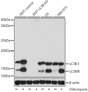
Western blot analysis of various lysates, using [KD Validated] LC3B Rabbit mAb (CAB19665) at 1:1000 dilution. 293T, C6 and NIH/3T3 cells were treated with Chloroquine (50 μM) at 37℃ for 20 hours. Secondary antibody: HRP-conjugated Goat anti-Rabbit IgG (H+L) (CABS014) at 1:10000 dilution. Lysates/proteins: 25μg per lane. Blocking buffer: 3% nonfat dry milk in TBST. Detection: ECL Basic Kit (AbGn00020). Exposure time: 5s.
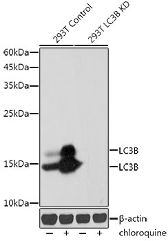
Western blot analysis of lysates from wild type(WT) and LC3B knockdown (KD) 293T cells, using [KD Validated] LC3B Rabbit mAb (CAB19665) at 1:1000 dilution. wild type(WT) and LC3B knockdown (KD) 293T cells were treated with Chloroquine (50 μM) at 37℃ for 20 hours. Secondary antibody: HRP-conjugated Goat anti-Rabbit IgG (H+L) (CABS014) at 1:10000 dilution. Lysates/proteins: 25μg per lane. Blocking buffer: 3% nonfat dry milk in TBST. Detection: ECL Basic Kit (AbGn00020). Exposure time: 30s.
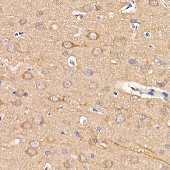
Immunohistochemistry analysis of paraffin-embedded Rat brain using [KD Validated] LC3B Rabbit mAb (CAB19665) at dilution of 1:100 (40x lens). High pressure antigen retrieval performed with 0.01M Citrate buffer (pH 6.0) prior to IHC staining.
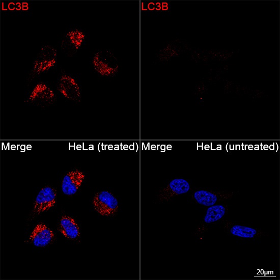
Confocal imaging of HeLa cells (treated with Chloroquine) and HeLa cells (untreated) using [KD Validated] LC3B Rabbit mAb (CAB19665, dilution 1:200) followed by a further incubation with Cy3 Goat Anti-Rabbit IgG (H+L) (CABS007, dilution 1:500) (Red). DAPI was used for nuclear staining (Blue). Objective: 100x.
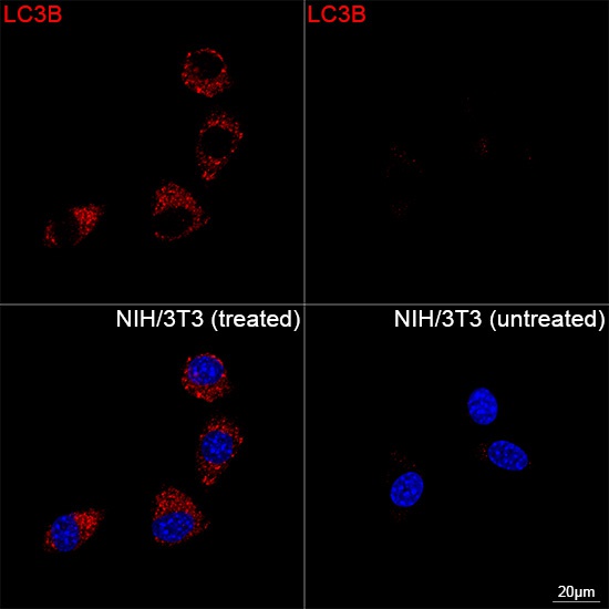
Confocal imaging of NIH/3T3 cells (treated with Chloroquine) and NIH/3T3 cells (untreated) using [KD Validated] LC3B Rabbit mAb (CAB19665, dilution 1:200) followed by a further incubation with Cy3 Goat Anti-Rabbit IgG (H+L) (CABS007, dilution 1:500) (Red). DAPI was used for nuclear staining (Blue). Objective: 100x.
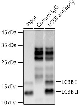
Immunoprecipitation analysis of 300 μg extracts from 293T cells using 3 μg [KD Validated] LC3B Rabbit mAb (CAB19665). Western blot was performed from the immunoprecipitate using LC3B antibody (CAB19665) at a dilution of 1:1000.