The [KO Validated] MEK1 Antibody (CAB12687) is a high-quality antibody developed for reliable detection and analysis of target proteins. This antibody, generated in rabbits, exhibits high reactivity with human samples and has been validated for use in Western blotting applications. By targeting the MAP2K1 protein, this antibody enables researchers to detect and analyze MAP2K1 expression in various cell types, making it ideal for studies in cell signaling, cancer research, and drug development.MAP2K1, also known as MEK1, plays a crucial role in transmitting signals from cell surface receptors to the nucleus, ultimately influencing cellular processes such as proliferation, differentiation, and survival.
This antibody is validated for use in WB, IHC-P, ELISA applications and has demonstrated reactivity against Human, Mouse, Rat samples.
Product Name:
[KO Validated] MEK1 Antibody
SKU:
CAB12687
Size:
20μL, 100μL
Reactivity:
Human, Mouse, Rat
Conjugate:
Unconjugated
Immunogen:
Synthetic peptide. This information is considered to be commercially sensitive.
Recommended starting concentration is 1 μg/mL. Please optimize the concentration based on your specific assay requirements.
Synonyms:
MEL, CFC3, MEK1, MKK1, MAPKK1, PRKMK1, K1
Positive Sample:
Jurkat, HeLa, A-431, MCF-7, Mouse Liver, Mouse Kidney, Rat Thymus
Cellular Localization:
Cytoplasm, Membrane, Nucleus, Peripheral Membrane Protein, Centrosome, Cytoskeleton, Microtubule Organizing Center, Spindle Pole Body.
Calculated MW:
43kDa
Observed MW:
45kDa
The protein encoded by this gene is a member of the dual specificity protein kinase family, which acts as a mitogen-activated protein (MAP) kinase kinase. MAP kinases, also known as extracellular signal-regulated kinases (ERKs), act as an integration point for multiple biochemical signals. This protein kinase lies upstream of MAP kinases and stimulates the enzymatic activity of MAP kinases upon wide variety of extra- and intracellular signals. As an essential component of MAP kinase signal transduction pathway, this kinase is involved in many cellular processes such as proliferation, differentiation, transcription regulation and development.
Purification Method
Affinity purification
Gene ID
5604
RRID
AB_2861674
Buffer Information
Store at -20℃. Avoid freeze / thaw cycles. Buffer: PBS with 0.01% thimerosal,50% glycerol,pH7.3.
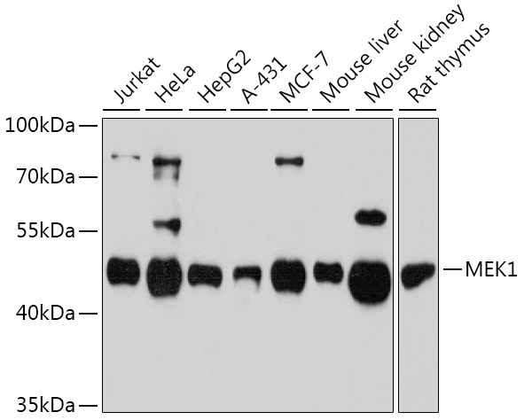
Western blot analysis of various lysates using [KO Validated] MEK1 Rabbit pAb (CAB12687) at 1:3000 dilution. Secondary antibody: HRP-conjugated Goat anti-Rabbit IgG (H+L) (CABS014) at 1:10000 dilution. Lysates/proteins: 25μg per lane. Blocking buffer: 3% nonfat dry milk in TBST. Detection: ECL Enhanced Kit (AbGn00021). Exposure time: 90s.
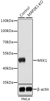
Western blot analysis of lysates from wild type (WT) and MEK1 knockout (KO) HeLa cells, using [KO Validated] MEK1 Rabbit pAb (CAB12687) at 1:1000 dilution. Secondary antibody: HRP-conjugated Goat anti-Rabbit IgG (H+L) (CABS014) at 1:10000 dilution. Lysates/proteins: 25μg per lane. Blocking buffer: 3% nonfat dry milk in TBST. Detection: ECL Basic Kit (AbGn00020). Exposure time: 5s.
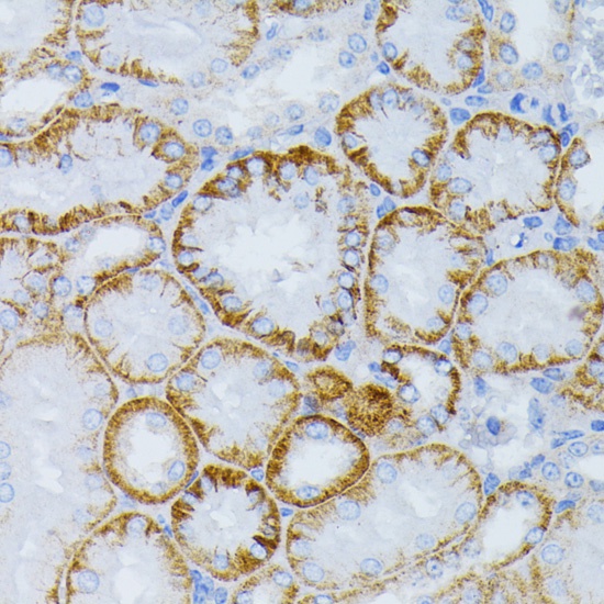
Immunohistochemistry analysis of paraffin-embedded Mouse kidney using [KO Validated] MEK1 Rabbit pAb (CAB12687) at dilution of 1:100 (40x lens). Microwave antigen retrieval performed with 0.01M PBS Buffer (pH 7.2) prior to IHC staining.