The [KO Validated] N-Cadherin Monoclonal Antibody (CAB19083) is a high-quality antibody developed for reliable detection and analysis of target proteins. This antibody, generated in rabbits, is highly specific for human samples and has been validated for use in applications such as Western blot and immunofluorescence.N-Cadherin is essential for maintaining tissue integrity and function, with dysregulation of its expression linked to various diseases including cancer and developmental disorders.
This antibody is validated for use in WB, IHC-P, ELISA, IF-P applications and has demonstrated reactivity against Human, Mouse, Rat samples.
Product Name:
[KO Validated] N-Cadherin Monoclonal Antibody
SKU:
CAB19083
Size:
20μL, 100μL
Reactivity:
Human, Mouse, Rat
Clone Number:
ARC0371
Conjugate:
Unconjugated
Immunogen:
Synthetic peptide. This information is considered to be commercially sensitive.
Recommended starting concentration is 1 μg/mL. Please optimize the concentration based on your specific assay requirements.
Synonyms:
CDHN, NCAD, ACOGS, ADHD8, CD325, ARVD14, CDw325, in
Positive Sample:
HeLa, C2C12, C6
Cellular Localization:
Cell Membrane, Single-Pass Type I Membrane Protein.
Calculated MW:
100kDa
Observed MW:
140kDa
This gene encodes a classical cadherin and member of the cadherin superfamily. Alternative splicing results in multiple transcript variants, at least one of which encodes a preproprotein is proteolytically processed to generate a calcium-dependent cell adhesion molecule and glycoprotein. This protein plays a role in the establishment of left-right asymmetry, development of the nervous system and the formation of cartilage and bone.
Purification Method
Affinity purification
Gene ID
1000
RRID
AB_2862575
Buffer Information
Store at -20℃. Avoid freeze / thaw cycles. Buffer: PBS containing 50% glycerol and 0.05% BSA, preserved with proclin300 or sodium azide, pH 7.3.
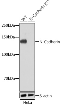
Western blot analysis of lysates from wild type (WT) and N-Cadherin knockout (KO) HeLa cells using [KO Validated] N-Cadherin Rabbit mAb (CAB19083) at 1:1000 dilution incubated overnight at 4℃. Secondary antibody: HRP-conjugated Goat anti-Rabbit IgG (H+L) (CABS014) at 1:10000 dilution. Lysates/proteins: 25μg per lane. Blocking buffer: 3% nonfat dry milk in TBST. Detection: ECL Basic Kit (AbGn00020). Exposure time: 1min.
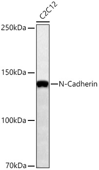
Western blot analysis of lysates from C2C12 cells using [KO Validated] N-Cadherin Rabbit mAb (CAB19083) at 1:1000 dilution incubated overnight at 4℃. Secondary antibody: HRP-conjugated Goat anti-Rabbit IgG (H+L) (CABS014) at 1:10000 dilution. Lysates/proteins: 25 μg per lane. Blocking buffer: 3% nonfat dry milk in TBST. Detection: ECL Basic Kit (AbGn00020). Exposure time: 60s.
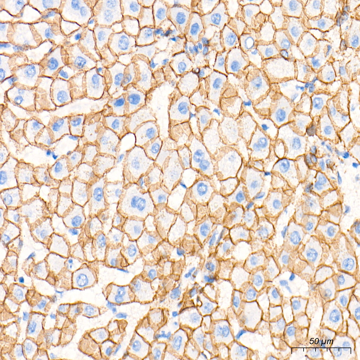
Immunohistochemistry analysis of paraffin-embedded Human liver tissue using [KO Validated] N-Cadherin Rabbit mAb (CAB19083) at a dilution of 1:2000 (40x lens). High pressure antigen retrieval performed with 0.01M Tris-EDTA Buffer (pH 9.0) prior to IHC staining.
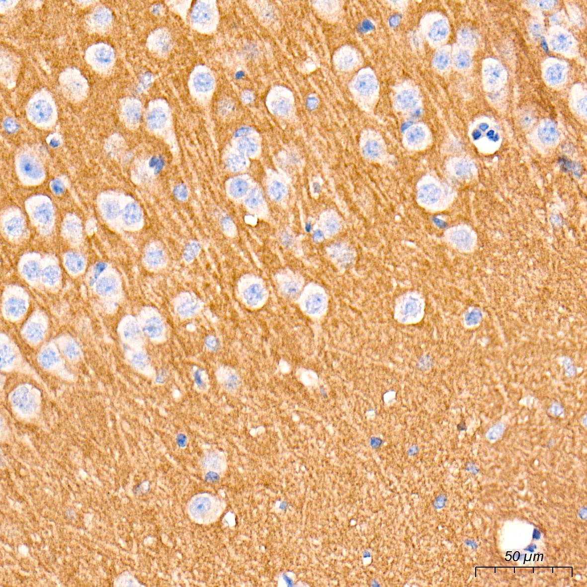
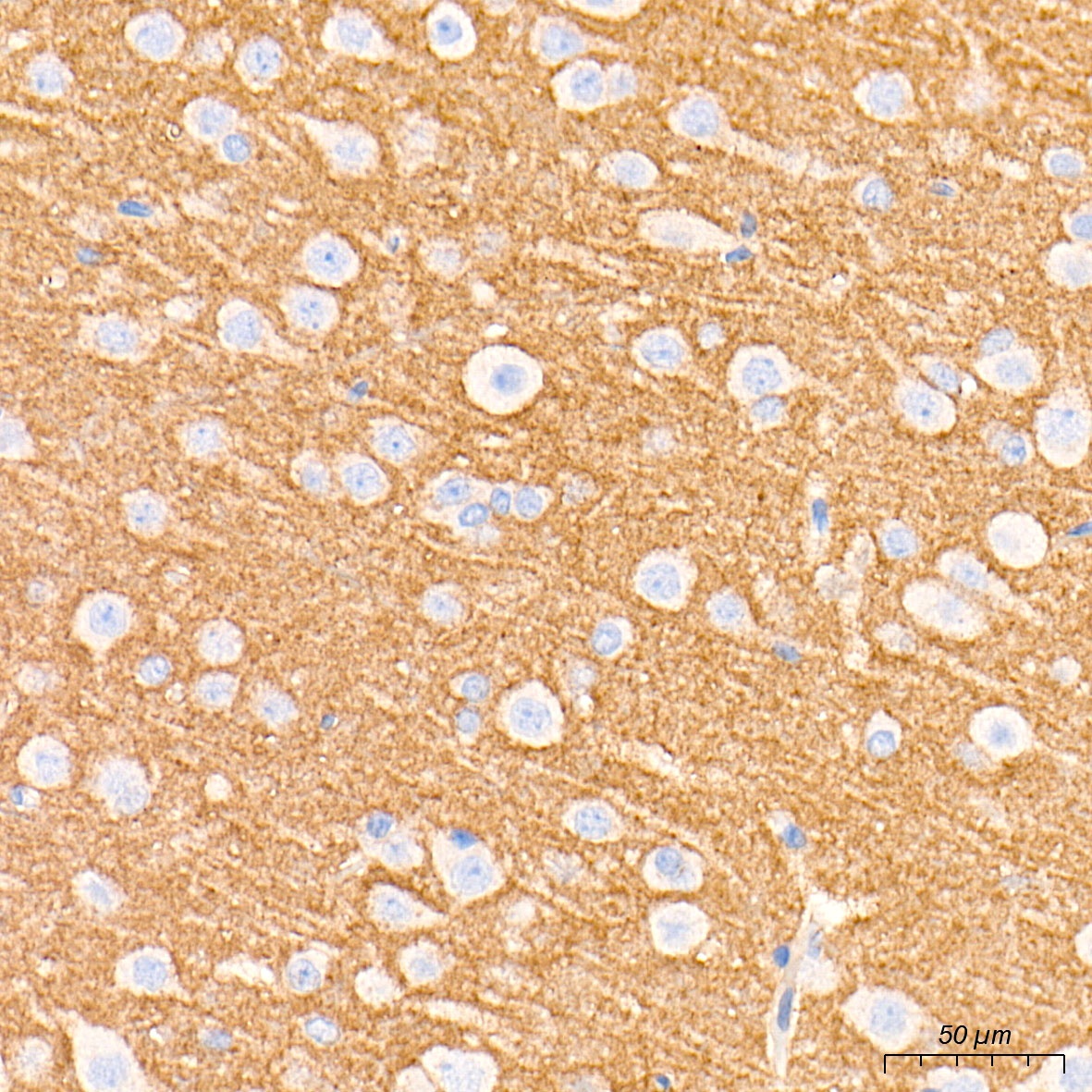
Immunohistochemistry analysis of paraffin-embedded Mouse brain tissue using [KO Validated] N-Cadherin Rabbit mAb (CAB19083) at a dilution of 1:2000 (40x lens). High pressure antigen retrieval performed with 0.01M Tris-EDTA Buffer (pH 9.0) prior to IHC staining.
Immunohistochemistry analysis of paraffin-embedded Rat brain tissue using [KO Validated] N-Cadherin Rabbit mAb (CAB19083) at a dilution of 1:2000 (40x lens). High pressure antigen retrieval performed with 0.01M Tris-EDTA Buffer (pH 9.0) prior to IHC staining.
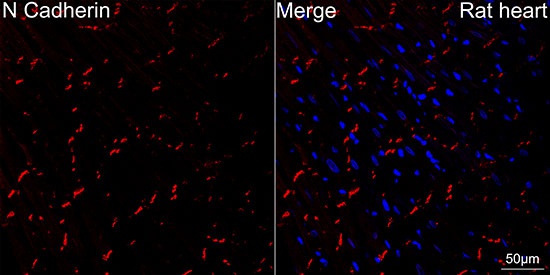
Confocal imaging of paraffin-embedded rat heart using [KO Validated] N-Cadherin Rabbit mAb (CAB19083, dilution 1:200) followed by a further incubation with Cy3 Goat Anti-Rabbit IgG (H+L) (CABS007, dilution 1:500) (Red). DAPI was used for nuclear staining (Blue). Objective: 40x. High pressure antigen retrieval performed with 0.01M Citrate Buffer(pH 6.0) prior to IF staining.
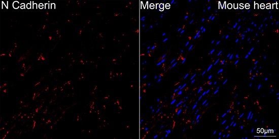
Confocal imaging of paraffin-embedded Mouse heart using [KO Validated] N-Cadherin Rabbit mAb (CAB19083, dilution 1:200) followed by a further incubation with Cy3 Goat Anti-Rabbit IgG (H+L) (CABS007, dilution 1:500) (Red). DAPI was used for nuclear staining (Blue). Objective: 40x. High pressure antigen retrieval performed with 0.01M Citrate Buffer(pH 6.0) prior to IF staining.