The [KO Validated] N-Cadherin Monoclonal Antibody (CAB19083) is a high-quality antibody developed for reliable detection and analysis of target proteins. This gene encodes a classical cadherin and member of the cadherin superfamily. Alternative splicing results in multiple transcript variants, at least one of which encodes a preproprotein is proteolytically processed to generate a calcium-dependent cell adhesion molecule and glycoprotein. This protein plays a role in the establishment of left-right asymmetry, development of the nervous system and the formation of cartilage and bone.
This antibody is validated for use in WB, IHC-P, ELISA, IF-P applications and has demonstrated reactivity against Human, Mouse, Rat samples.
Product Name:
[KO Validated] N-Cadherin Monoclonal Antibody
SKU:
CAB19083
Size:
100μL, 20μL
Reactivity:
Human, Mouse, Rat
Clone Number:
ARC0371
Conjugate:
Unconjugated
Immunogen:
Synthetic peptide. This information is considered to be commercially sensitive.
Tested Applications:
WBIHC-PELISAIF-P
Recommended Dilution:
WB
1:1000 - 1:2000
IF-P
1:50 - 1:200
IHC-P
1:1000 - 1:4000
ELISA
Recommended starting concentration is 1 μg/mL. Please optimize the concentration based on your specific assay requirements.
Synonyms:
CDHN, NCAD, ACOGS, ADHD8, CD325, ARVD14, CDw325, in
Positive Sample:
HeLa, C2C12, C6
Cellular Localization:
Cell Membrane, Single-Pass Type I Membrane Protein.
Calculated MW:
100 kDa
Observed MW:
140 kDa
This gene encodes a classical cadherin and member of the cadherin superfamily. Alternative splicing results in multiple transcript variants, at least one of which encodes a preproprotein is proteolytically processed to generate a calcium-dependent cell adhesion molecule and glycoprotein. This protein plays a role in the establishment of left-right asymmetry, development of the nervous system and the formation of cartilage and bone.
Purification Method
Affinity purification
Gene ID
1000
RRID
AB_2862575
Buffer Information
Store at -20℃. Avoid freeze / thaw cycles. Buffer: PBS containing 50% glycerol and 0.05% BSA, preserved with proclin300 or sodium azide, pH 7.3.
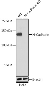
Western blot analysis of lysates from wild type (WT) and N-Cadherin knockout (KO) HeLa cells using [KO Validated] N-Cadherin Rabbit mAb (CAB19083) at 1:1000 dilution incubated overnight at 4℃. Secondary antibody: HRP-conjugated Goat anti-Rabbit IgG (H+L) (AS014) at 1:10000 dilution. Lysates/proteins: 25μg per lane. Blocking buffer: 3% nonfat dry milk in TBST. Detection: ECL Basic Kit (AbGn00020). Exposure time: 1min.
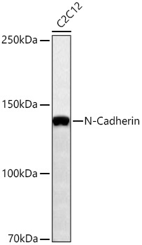
Western blot analysis of lysates from C2C12 cells using [KO Validated] N-Cadherin Rabbit mAb (CAB19083) at 1:1000 dilution incubated overnight at 4℃. Secondary antibody: HRP-conjugated Goat anti-Rabbit IgG (H+L) (AS014) at 1:10000 dilution. Lysates/proteins: 25 μg per lane. Blocking buffer: 3% nonfat dry milk in TBST. Detection: ECL Basic Kit (AbGn00020). Exposure time: 60s.
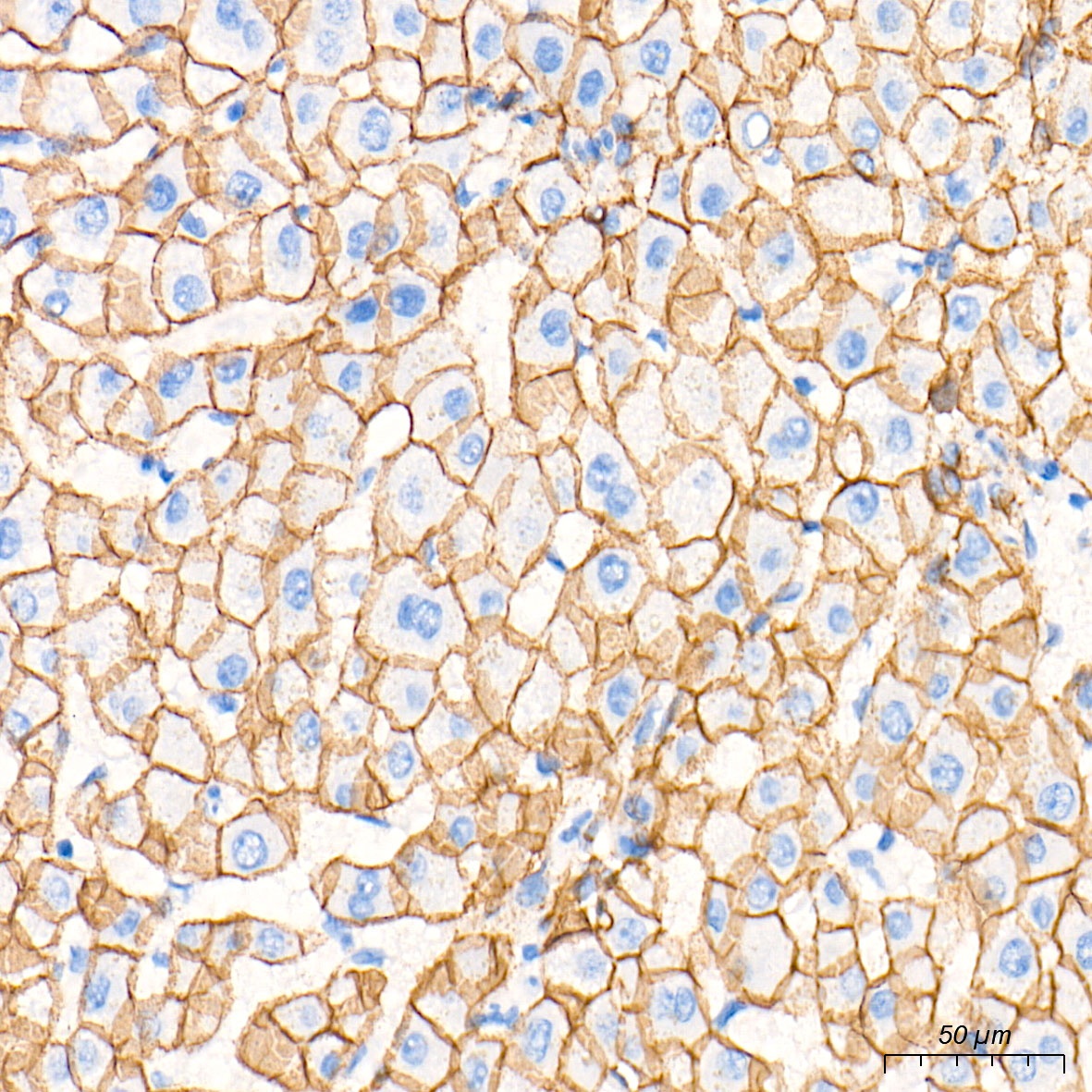
Immunohistochemistry analysis of paraffin-embedded Human liver tissue using [KO Validated] N-Cadherin Rabbit mAb (CAB19083) at a dilution of 1:2000 (40x lens). High pressure antigen retrieval performed with 0.01M Tris-EDTA Buffer (pH 9.0) prior to IHC staining.
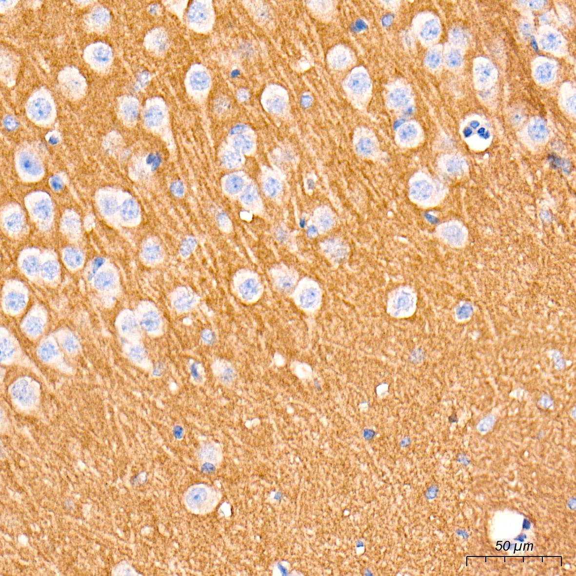
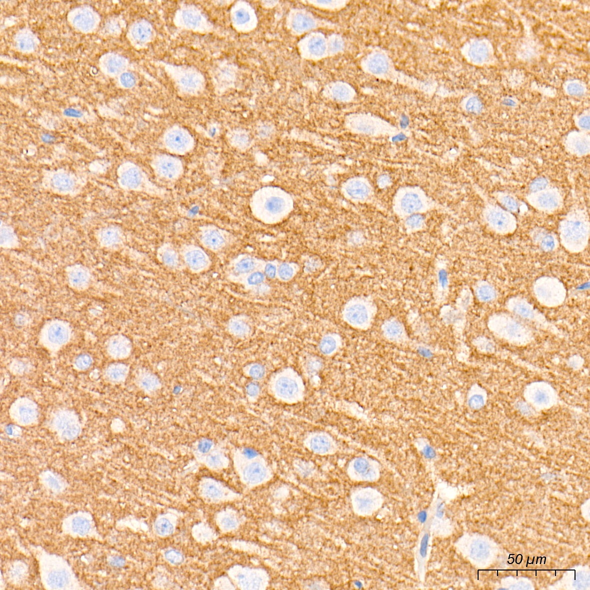
Immunohistochemistry analysis of paraffin-embedded Mouse brain tissue using [KO Validated] N-Cadherin Rabbit mAb (CAB19083) at a dilution of 1:2000 (40x lens). High pressure antigen retrieval performed with 0.01M Tris-EDTA Buffer (pH 9.0) prior to IHC staining.
Immunohistochemistry analysis of paraffin-embedded Rat brain tissue using [KO Validated] N-Cadherin Rabbit mAb (CAB19083) at a dilution of 1:2000 (40x lens). High pressure antigen retrieval performed with 0.01M Tris-EDTA Buffer (pH 9.0) prior to IHC staining.
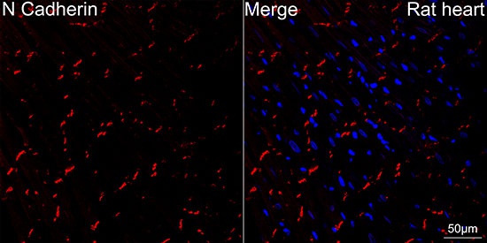
Confocal imaging of paraffin-embedded rat heart using [KO Validated] N-Cadherin Rabbit mAb (CAB19083, dilution 1:200) followed by a further incubation with Cy3 Goat Anti-Rabbit IgG (H+L) (AS007, dilution 1:500) (Red). DAPI was used for nuclear staining (Blue). Objective: 40x. High pressure antigen retrieval performed with 0.01M Citrate Buffer(pH 6.0) prior to IF staining.
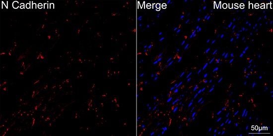
Confocal imaging of paraffin-embedded Mouse heart using [KO Validated] N-Cadherin Rabbit mAb (CAB19083, dilution 1:200) followed by a further incubation with Cy3 Goat Anti-Rabbit IgG (H+L) (AS007, dilution 1:500) (Red). DAPI was used for nuclear staining (Blue). Objective: 40x. High pressure antigen retrieval performed with 0.01M Citrate Buffer(pH 6.0) prior to IF staining.