The [KO Validated] SAMHD1 Antibody (CAB7794) is a high-quality antibody developed for reliable detection and analysis of target proteins. This polyclonal antibody, generated in rabbits, is specifically reactive with human samples and has been validated for use in various applications, including Western blotting.SAMHD1, known for its role in inhibiting HIV-1 replication, has also been implicated in DNA repair and modulation of cellular dNTP levels. By targeting the SAMHD1 protein, this antibody enables researchers to investigate its function in different cell types and conditions, making it ideal for studies in virology, immunology, and cancer research.
This antibody is validated for use in WB, IHC-P, IP, ELISA applications and has demonstrated reactivity against Human, Mouse, Rat samples.
Product Name:
[KO Validated] SAMHD1 Antibody
SKU:
CAB7794
Size:
20μL, 100μL
Reactivity:
Human, Mouse, Rat
Conjugate:
Unconjugated
Immunogen:
Recombinant protein (or fragment).This information is considered to be commercially sensitive.
0.5μg-4μg antibody for 200μg-400μg extracts of whole cells
ELISA
Recommended starting concentration is 1 μg/mL. Please optimize the concentration based on your specific assay requirements.
Synonyms:
DCIP, CHBL2, HDDC1, MOP-5, SBBI88, hSAMHD1, D1
Positive Sample:
HepG2, HeLa, MCF-7, Mouse spleen
Cellular Localization:
Nucleus.
Calculated MW:
72kDa
Observed MW:
72kDa
This gene may play a role in regulation of the innate immune response. The encoded protein is upregulated in response to viral infection and may be involved in mediation of tumor necrosis factor-alpha proinflammatory responses. Mutations in this gene have been associated with Aicardi-Goutieres syndrome.
Purification Method
Affinity purification
Gene ID
25939
RRID
AB_2863562
Buffer Information
Store at -20℃. Avoid freeze / thaw cycles. Buffer: PBS containing 50% glycerol, preserved with proclin300 or sodium azide, pH 7.3.
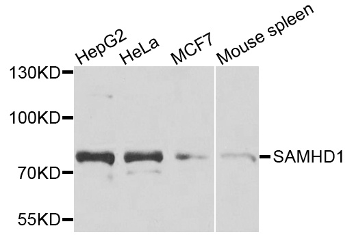
Western blot analysis of various lysates using [KO Validated] SAMHD1 Rabbit pAb (CAB7794) at 1:1000 dilution. Secondary antibody: HRP-conjugated Goat anti-Rabbit IgG (H+L) (CABS014) at 1:10000 dilution. Lysates/proteins: 25μg per lane. Blocking buffer: 3% nonfat dry milk in TBST. Detection: ECL Basic Kit (AbGn00020). Exposure time: 90s.
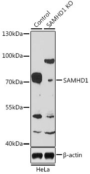
Western blot analysis of lysates from wild type (WT) and SAMHD1 knockout (KO) HeLa cells, using [KO Validated] SAMHD1 Rabbit pAb (CAB7794) at 1:1000 dilution. Secondary antibody: HRP-conjugated Goat anti-Rabbit IgG (H+L) (CABS014) at 1:10000 dilution. Lysates/proteins: 25μg per lane. Blocking buffer: 3% nonfat dry milk in TBST. Detection: ECL Basic Kit (AbGn00020). Exposure time: 3min.
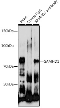
Immunoprecipitation analysis of 200 μg extracts of HeLa cells using 3 μg SAMHD1 antibody (CAB7794). Western blot was performed from the immunoprecipitate using SAMHD1 antibody (CAB7794) at a dilution of 1:1000.